Bipolar disorder (BD) is a chronic mental illness characterized by changes in mood
that alternate between mania and hypomania or between depression and mixed states,
often associated with functional impairment. Although effective pharmacological and
non-pharmacological treatments are available, several patients with BD remain symptomatic.
The advance in the understanding of the neurobiology underlying BD could help in the
identification of new therapeutic targets as well as biomarkers for early detection,
prognosis, and response to treatment in BD. In this review, we discuss genetic, epigenetic,
molecular, physiological and neuroimaging findings associated with the neurobiology
of BD. Despite the advances in the pathophysiological knowledge of BD, the diagnosis
and management of the disease are still essentially clinical. Given the complexity
of the brain and the close relationship between environmental exposure and brain function,
initiatives that incorporate genetic, epigenetic, molecular, physiological, clinical,
environmental data, and brain imaging are necessary to produce information that can
be translated into prevention and better outcomes for patients with BD.
Received: September 27 2019; Accepted: December 27 2019
INTRODUCTION
Bipolar disorder (BD) is a severe and chronic psychiatric illness that affects approximately
1-4% of the world population.1 It is characterized by changes in mood that alternate between mania and hypomania
or between depression and mixed states, often associated with functional impairment.1-3 Although depressive symptoms typically predominate in individuals with BD, the clinical
diagnostic hallmark is the presence of manic or hypomanic episodes.4 During episodes of mania or hypomania, the patient may present symptoms related to
elevated mood, including euphoria, feelings of greatness, hyperactivity, increased
sexual activity, decreased need for sleep, risky behaviors, irritability, and aggression.
Conversely, episodes of depression are characterized by anhedonia, sadness, vegetative
symptoms, and psychomotor retardation. Mixed episodes manifest as simultaneous states
of mania and depression.4 About 14-59% of the individuals with BD report suicidal ideation5; 25 to 50% attempt suicide, and almost 20% die due to suicide.6 Moreover, BD is also associated with increased risk of mortality by other medical
conditions, such as cardiovascular disease, diabetes mellitus, external causes of
injuries, and respiratory diseases. Life expectancy is decreased by 9 years on average
in individuals with BD as compared with the general population.7
Despite the demonstrated efficacy of several drugs to treat BD, lithium (Li) is the
only drug considered as a mood stabilizer by the Food and Drug Administration (FDA).
More than 70 years have passed since this salt was first proposed as a mood stabilizer.
Li is effectively used in BD for maintenance and treatment of acute mania episodes;
however, it has a modest antidepressant action.8 Anticonvulsants such as valproate (VPA) and carbamazepine are also used to treat
BD. Additionally, some typical (e.g., haloperidol and chlorpromazine) and atypical
antipsychotics (e.g., aripiprazole, clozapine, olanzapine, quetiapine, risperidone,
ziprasidone, and asenapine) have also shown efficacy.9-11 Of note, the treatment of depression in BD is particularly challenging, as several
drugs used for treating depressive symptoms, such as selective serotonin reuptake
inhibitors (SSRI), can induce a switch to hypomania or mania. Thus, these drugs are
usually combined with Li for acute treatment and maintenance of bipolar depression.12 It is noteworthy that patients with BD commonly face residual mood symptoms, cognitive
and functional impairment, psychosocial disability, and decreased quality of life
even with the best treatment available.13,14
BD has been suggested as a progressive condition, and the delay in diagnosis as well
as inappropriate treatment can result in repeated mood episodes, persistent subthreshold
symptoms, development of co-morbidities, and progression of the disease with cognitive
impairment and functional decline.15-17 The term "neuroprogression" has thus been conceptualized as the pathological rewiring
of the brain that takes place in parallel with the functional and clinical deterioration
that may occur in the course of BD; according to this concept, different stages of
BD are associated with distinct neurobiological underpinnings.18-23 Indeed, studies have shown that structural brain changes and cognitive deficits are
not consistently found at illness onset, and appear to become more evident with chronicity
and recurring episodes.24-27 Moreover, neuroprogression seems to be related to the cumulative effects of immune
dysfunction, enhanced oxidative stress, neurotrophic support breakdown, mitochondrial
dysfunction, and impairment of cellular resilience.20,21,28,29 Neuroprogression has been also associated with lower responsiveness to treatment,
especially with Li and cognitive behavioral therapy.30-32 In this sense, Post et al.33 have suggested that multiple episodes may lead to permanent alterations in neuronal
activity, which may translate into greater liability to relapse and poorer treatment
response. Of note, such a progressive feature does not seem to be present across all
patients, with some not experiencing as much cognitive or functional impairment as
others.34 This poses the particular challenge of identifying specific subgroups of susceptible
patients, which may require a deeper understanding of the biological basis of BD.
Although numerous factors and mechanisms have been proposed to explain the pathophysiology
of BD, its definitive etiopathology remains unknown. Nonetheless, it is believed that
the etiology of BD involves an interaction between multiple genetic, neurochemical,
and environmental factors (Figure 1).35 Not only would a deep understanding of the neurobiology of BD support the discovery
of new targets for effective pharmacological and non-pharmacological therapies, it
would also facilitate the identification of biomarkers for diagnosis, prognosis, and
response to specific treatments. The last years have witnessed great progress in the
understanding of BD pathophysiology. For instance, several lines of thought have led
us to believe that BD is associated with neuroprogression, and that over time recurrence
may exacerbate subsequent episodes, which are accompanied by functional and cognitive
impairment.20,36-38 An underlying hypothesis is that changes in neuroplasticity, with a decrease in neurotrophic
factors combined with mitochondrial dysfunction, oxidative stress, inflammation, circadian
rhythm abnormalities, and biological aging acceleration, could be associated with
the worsening of mood episodes, refractoriness to treatment, cognitive impairment,
and functional disability.20 This article seeks to discuss studies addressing the neurobiological mechanisms of
BD, providing an overview of genetic components, major signaling pathways, biochemical
changes, and neuroimaging findings associated with this disorder.
Figure 1. Etiology of bipolar disorders. The cause of bipolar disorder (BD) is still unknown.
However, it has been established that the dynamic interplay between genetic, neurochemical,
and environmental factors plays a role in the onset and progression of BD.
Figure 1. Etiology of bipolar disorders. The cause of bipolar disorder (BD) is still
unknown. However, it has been established that the dynamic interplay between genetic,
neurochemical, and environmental factors plays a role in the onset and progression
of BD.
GENETICS AND EPIGENETICS OF BIPOLAR DISORDER
Evidence suggests that BD presents a very strong genetic component, with twin studies
showing heritability rates (i.e., the extent to which the disorder can be explained
by genetic factors) as high as 70-80%.39,40 The strength of this genetic component is also supported by the increased risk of
BD noted in first-degree relatives of patients, including offspring.41 The risk of BD is significantly higher in children of parents diagnosed with BD vs.
offspring of control parents42,43; indeed, a plethora of signs and symptoms may accompany such familial risk even in
the absence of a full-blown diagnosis of BD.44-48 This familial aggregation suggests a potential relevance of both inherited genes,
which can be tested by molecular genetic analyses, and the "inherited" familial environment,
which is also known to be potentially impaired in the face of parental psychopathology.49,50
In the search for relevant BD-related genes, several genome-wide association studies
(GWASs) conducted in the past years have identified multiple loci with a small effect that may account for heritability,51-62 including 30 recently discovered loci encoding ion channels, neurotransmitter transporters, and synaptic components.63 None of these single nucleotide polymorphisms (SNPs), however, has been shown to
present high penetrance or a large effect size, a finding that is consistent with
the complex multifactorial model that is believed to underlie the genetics of BD and
other psychiatric disorders. Moreover, the difference in heritability rate calculated
through twin studies (70-80%) and that calculated based on molecular genetics findings
(approximately 30%) is quite large, suggesting a role of other markers or mechanisms.64 These may include gene-by-gene interactions, rare genetic markers (current GWASs
only assess "common" alterations), gene-by-environment interactions, and epigenetic
markers.
Epigenetic mechanisms encompass several pathways that can mediate gene-by-environment
interactions and modulate gene expression and activity without altering the DNA sequence.
These include, among others, DNA methylation, histone modifications, chromatin remodeling,
and the actions of noncoding RNAs. Several of these have been suggested to play significant
roles in BD pathophysiology, especially DNA methylation.65-67 In addition, several epigenetic alterations that may underlie risk/resilience mechanisms
have been described in youth at high risk of BD, such as offspring of BD parents.68,69 Specifically, DNA methylation is a fairly stable epigenetic alteration that has been
implicated in BD pathogenesis and progression,69-71 as well as in the mechanisms of action of several drugs used to treat patients.65,72-74 DNA methylation in promoters is typically associated with the repression of gene
expression and may underlie at least some of the transcriptomic changes reported for
BD in multiple tissues.75-79 Moreover, several preclinical and clinical studies have shown that early-life traumatic
experiences can induce stable methylation alterations that persist into adulthood,
suggesting epigenetic-based alterations as mediators of the clinical effects of early
adversity.80-82
Interestingly, one of the mechanisms by which methylomes have been shown to interfere
with BD phenotype is the modulation of biological aging processes. Patients with BD
have been shown to present several clinical markers that are suggestive of premature
aging, including a higher rate of age-related conditions and faster cognitive decline,
with previous reports suggesting shortened telomere length compared to controls.83-85 More recently, a marker of biological aging based solely on DNA methylation levels
("epigenetic age" or "DNA methylation age")86 has been used to explore the aging processes in BD, and the results suggest accelerated
epigenetic aging in the blood and brain of BD patients compared to controls.71,87 Recent evidence that the epigenetic clock can be pharmacologically reversed in humans88 offers an interesting treatment possibility for the modulation of premature aging
in BD.
In addition to DNA methylation, noncoding RNAs have also deserved much attention,
including gene expression modulators such as microRNAs, long noncoding RNAs, and others.89 Several alterations in the levels of microRNAs and long noncoding RNAs have been
detected in BD samples, some of which have been validated by independent studies.90,91 Recent studies have also suggested that noncoding RNAs may function as clinically
relevant peripheral biomarkers when assessed from neuron-derived extracellular vesicles,
which are currently being investigated in BD patients.92,93 Altogether, these studies suggest an important role for noncoding RNAs in BD pathophysiology,
and also their potential as an important diagnostic and prognostic tool.
CHANGES IN NEUROPLASTICITY AND NEUROTROPHIC SIGNALING
Neurotrophic factors comprise a group of proteins responsible for regulating neuronal
survival processes, neuronal growth, synaptic formation, and cellular plasticity at
the central and peripheral nervous systems. Nerve growth factor (NGF), brain-derived
neurotrophic factor (BDNF), glial-derived neurotrophic factor (GDNF), neurotrophin-3
(NT-3), and neurotrophin-4 (NT-4) are the most common neurotrophic factors studied
in psychiatric disorders. However, BDNF is undoubtedly the most studied neurotrophin
in BD. Neurotrophins are a specific type of neurotrophic factor, and members of this
family include BDNF, NGF, NT-3, and NT-4. Neurotrophins bind and activate a specific
family of tyrosine kinase (Trk) receptors, thus promoting modulation of the central
nervous system (CNS). There are three specific Trk receptors: NGF binds to TrkA; BDNF
and NT-4 bind to TrkB; and NT-3 binds to the TrkC receptor.94
The strong interest in BDNF and other neurotrophins in BD was triggered by the discovery
that antidepressants and mood stabilizers could act on these molecules, modulating
their signaling pathways. Preclinical studies have shown that the chronic use of antidepressants
and mood stabilizers, such as Li and VPA, can increase NGF, BDNF, and GDNF levels
in the rat brain.95-98 Currently, a range of studies show decreased levels of BDNF and its receptor TrkB
in both blood and brain of patients with BD.99-103 Additionally, a BDNF gene polymorphism that replaces a valine for a methionine (i.e.,
valine replacing methionine) at codon 66 (Val66Met) has been repeatedly associated
with BD.104 A previous study also demonstrated decreased plasma NGF levels in patients with BD.105 Another clinical study showed that NT-3 and NT-4 are elevated in the depressive phase
of BD,106 possibly indicating an attempt by the organism to defend itself against cellular
stress. In turn, Barbosa et al.107 demonstrated decreased NT-3 and NT-4 levels in the manic phase of BD. Given that
neurotrophins are essential for neuronal function and survival, it is assumed that
the viability of nerve cells can be affected by a persistent reduction of these molecules
in the nervous system.
As in clinical studies, preclinical research has demonstrated that amphetamine (AMPH)
or ouabain-induced manic behaviors in rats decrease BDNF, NGF, and GDNF levels in
the brain of animals.95,98 Additionally, decreased levels of BDNF, NGF, and GDNF followed depressive-like behaviors
in an animal model of depression induced by maternal deprivation or chronic mild stress.108 A previous preclinical study suggested that neuroadaptations induced by chronic administration
of dextroamphetamine (d-AMPH) might mimic neuroprogression in BD, because the brain
is primed for both the manic and depressive episodes which are characteristic of BD.
In that study, the authors showed that d-AMPH withdrawal induces depressive- and anxious-like
behaviors in rats, as well as a sensitization to manic-like behaviors. In line with
this, d-AMPH sensitization decreased the levels of BDNF, NGF, and GDNF and increased
the levels of NT-3 and NT-4/5 in the brain of rats.109
Together, these clinical and preclinical studies suggest changes in neurotrophic factors
as an attractive molecular mechanism to explain the decreased cellular plasticity
observed in BD, along with other mechanisms described in detail in the next sections.
The hypothesis is that changes in neuroplasticity, including alterations in neurotrophic
signaling, could be associated with brain damage, which in turn worsens mood episodes
and ultimately induces cognitive and functioning deficits in patients with BD.
MITOCHONDRIAL DYSFUNCTION AND OXIDATIVE STRESS
Mitochondria are organelles that are responsible, both directly and indirectly, for
cellular functions such as energy production; they also function as sources of cellular
growth substrates and play crucial roles in oxidative/nitrosative stress, cell resilience
and cell death.110-112 In the brain, mitochondria are critical for the modulation of neuronal activity,
short- and long-term neuronal plasticity, cellular resilience, and behavioral adaptations,
mainly through actions on long-term potentiation, the hallmark process of learning
and memory.112-115 For more than 50 years, multiple studies have highlighted mitochondrial dysfunction
as a common pathway in BD pathophysiology, triggered by mechanisms such as impaired
oxidative phosphorylation, shift to glycolytic production of energy, general decrease
in energy, and abnormalities in the morphology and intracellular distribution of mitochondria.116 The "mitochondrial hypothesis" suggests that BD is triggered, at least in part, by
mitochondrial dysfunction, which can be intimately linked to a wide range of processes
associated with treatment outcomes and disease progression or severity, including
inflammation, oxidative stress, stress response systems, and accelerated aging.
A considerable number of studies in the literature has shown an increase in lactate
levels and a reduction in intracellular pH (ipH) in the brain of patients with BD,
possibly indicating altered mitochondrial function and a glycolytic shift consistent
with impaired mitochondrial metabolism in BD.116-120 Glycolysis-only adenosine triphosphate (ATP) production has been shown to be powerless
to maintain normal levels of Na+/K+-ATPase activity in neurons, inducing a large calcium influx into neurons, followed
by glutamate excitotoxicity and neuronal apoptosis, both of which play a central role
in neurodegeneration.121 Mallakh et al.122 propose that both manic and depressed states in BD could be caused by Na+/K+-ATPase dysfunction - a small reduction in Na+/K+-ATPase activity in the brain would lead to a hyperexcitable state (mania) by bringing
the resting potential of neurons closer to the threshold for activation and increasing
the duration of neurotransmitter release; in turn, a further reduction in Na+/K+-ATPase function would bring the resting potential even closer to the threshold for
activation, decreasing the amplitude of the action potential and resulting in inhibition
of neurotransmitter release (depression).122
An additional link between mitochondrial dysfunction and BD is supported by data demonstrating
that patients with BD present significantly lower levels of phosphocreatine (PCr)
(a high-energy compound) and adenosine diphosphate (ADP) and decreased PCr peak area
percentage, suggesting regional hypometabolism in all three mood states.123 Additionally, the PCr peak area percentage was observed to be decreased in the left
frontal lobe of patients during the depressive state, as well as in the right frontal
lobe during manic/euthymic states.124,125 Moreover, previous studies have shown that patients with BD present lower levels
of N-acetyl-aspartate (NAA), which is thought to represent a surrogate marker for
impaired mitochondria, along with a negative correlation between NAA/creatine + PCr
or NAA levels and illness duration.126-129 Taken together, these studies provide indirect evidence that mitochondrial dysfunction
may play a role in illness progression.20
Several clinical and animal studies have reported alterations in various energetic
metabolism components, including downregulation of nuclear mRNA molecules and proteins
involved in the Krebs cycle, electron transport chain (ETC) I-IV complexes, and creatine
kinase, as well as a marked decrease in the activity of ETC complexes and Krebs cycle
enzymes.130-143 Together, these studies suggest a reduced ability to oxidize NADH and FADH and transfer
electrons to ubiquinone in BD, resulting in reactive oxygen species (ROS) production
through an increase in the rate of electron leakage by ETC complexes.
More recent attention has focused on the maintenance of a healthy mitochondrial pool,
which is critically regulated by the dynamics and turnover of the mitochondrial population.
A damaged mitochondrion may segregate its damaged components into subcompartments
and divide, whereas a healthy mitochondrion with a potential healthier membrane will
continue to participate in fusion and fission cycles. Depolarized damaged mitochondria
are often degraded through mitophagy via the PTEN-induced kinase 1 (PINK1)-Parkin
pathway.144 Thus, these dynamic processes of mitochondrial fusion, fission, transport, and turnover
enable recruitment of healthy mitochondria to subcellular compartments with high demands
for ATP; disruptions in any of these processes will lead to mitochondrial pathology,
cellular dysfunction, and neurological defects.145 Previous research findings into mitochondrial morphology have shown that prefrontal
cortex neurons of the postmortem brain from patients with BD and peripheral cells
from living BD patients display morphological abnormalities (more mitochondria of
smaller size) and an abnormal pattern of clumping and marginalization in the intracellular
distribution of mitochondria.146 By drawing on the concept of mitochondrial quality control, recent studies have demonstrated
that patients with BD present an imbalance in mitochondrial fission and fusion toward
fission, followed by a decrease in the levels of mitophagy proteins and increase in
caspase-3 protein levels in peripheral blood mononuclear cells, suggesting that, in
patients with BD, the number of damaged mitochondria exceeds the capacity of mitophagy,
and apoptosis becomes the dominant pathway to minimize tissue damage.147,148
Moreover, numerous studies have provided evidence of increased ROS production and
oxidative stress in patients with BD. Replicated evidence has documented alterations
in multiple aspects of oxidative stress regulation, including the production of ROS
and reduced antioxidant capacity. Meta-analyses have shown significantly greater levels
of lipid peroxidation markers, DNA/RNA damage, and nitric oxide in BD.149,150 However, mixed results regarding two of the primary antioxidant enzymes, glutathione
peroxidase (GPx) and superoxide dismutase (SOD), have been reported in BD, followed
by a meta-analysis that did not reach statistical significance for the comparison
of GPx and SOD activity levels in individuals with BD vs. controls.149 Conversely, a recent study by Das et al.151 showed that, although glutathione levels did not differ in BD and controls, there
was a positive, BD-specific correlation between lactate and glutathione levels, indicating
a physiological association between the antioxidant system and mitochondrial dysfunction.
Several investigators have reported that excessive oxidative stress in pathological
conditions induces point mutations and may result in large deletions of mitochondrial
DNA (mtDNA) due to restricted DNA repair ability and the absence of histones in mitochondria.
Regarding BD, there is an inconsistency in mtDNA copy number (mtDNAcn) studies that
could be attributed to the diversity in clinical features, tissue types, and ethnicity.71,152-155 A recent meta-analysis identified no significant differences in mtDNAcn exhibited
by BD patients or controls, while an Asian-specific meta-analysis for BD-mtDNAcn studies
revealed significantly lower mtDNAcn in patients with BD, with a low level of heterogeneity.155 Moreover, mtDNAcn was inversely correlated with the number of relapses of manic episodes.154 A possible mechanism for the lower levels of mtDNAcn could be related to DNA polymerase
gamma (POLG) dysfunction due to oxidative stress. In fact, recent studies have demonstrated
that individuals with BD present downregulation of POLG, the replicative polymerase
responsible for maintaining mtDNA, as well as downregulation of the DNA repair enzyme,
8-oxoguanine-DNA glycosylase 1 (OGG1).156,157
The accumulating evidence reviewed above delineates multifaceted mitochondrial dysfunction
as a pathological factor in BD. Moreover, mitochondrial dysfunction, apoptosis, and
non-methylated mtDNA can activate Toll-like receptors and lead to spontaneous inflammasome
activation, stimulating cytokine production and inducing rapid activation of the immune
system.
IMMUNE-INFLAMMATORY IMBALANCE AND KYNURENINE PATHWAY
Immune dysfunction in BD is supported by pre-clinical and clinical evidence showing
elevated levels of pro-inflammatory cytokines, including interleukin-4 (IL-4), interleukin-1beta
(IL-1ß), interleukin-6 (IL-6), tumor necrosis factor (TNF)-alpha, soluble interleukin-2
receptor (sIL-2R), and soluble receptor of TNF-type 1 (STNFR1), among others, in patients
compared to controls.158-164 A recent systematic review has suggested an important role for acute inflammatory
response during mania and depression, with the elevation in proinflammatory cytokines
seemingly restored after remission of symptoms.118 In addition, further findings in BD have also described significant negative associations
between inflammatory markers and general cognitive abilities, as well as neuroanatomical
alterations.165-169 However, another systematic review claims that an absolute conclusion cannot be reached
regarding the presence of neuroinflammation in BD, since the findings are not consistent.170
Several different mechanisms have been proposed to explain the role of immune dysfunction
in BD,171 including changes in blood-brain barrier, cell death-induced release of damage-associate
molecular patterns with consequent immune activation, genetic mechanisms, dysfunction
of the gut-brain axis, and a role of the kynurenine pathway. Activation of the kynurenine
pathway by cytokines such as interferon-gamma (IFN-?) and TNF-a is described as one
of several contributors to psychiatric pathogeneses. Kynurenine metabolites mediate
immune-inflammation and neurodegeneration and can lead to neurotoxicity and impaired
neurotransmission. The enzyme indoleamine 2,3-dioxygenase (IDO-1) converts tryptophan
into kynurenine and is activated by inflammatory cytokines. Kynurenine is then metabolized
into hydroxykynurenine and quinolinic acid in microglia, and into kynurenic acid (KYNA)
in astrocytes. Hydroxykynurenine and quinolinic acid are N-methyl-D-aspartic acid
(NDMA) agonists and increase the production of free radicals, while KYNA, an N-methyl-D-aspartate
(NMDA) receptor antagonist, has neuroprotective effects but also leads to cognitive
deficits and psychosis when in higher levels.172-174
A previous study showed an imbalance toward the neurotoxicity path derived from kynurenine
metabolism, as evidenced by decreased levels of KYNA, in addition to increased hydroxykynurenine/kynurenine
ratio in BD compared with healthy controls.175 The higher hydroxykynurenine levels may correlate to cognitive dysfunction not only
in the acute phases of BD, but also in euthymic periods.27 Recent evidence shows that KYNA levels in cerebrospinal fluid represent a biomarker
for psychotic episodes in BD. This higher risk of developing psychosis may be explained
by enhanced dopaminergic transmission due to increased brain KYNA concentrations.176 Conversely, decreased peripheral KYNA and unchanged KYNA levels were observed in
the CNS in BD patients during the depressive phase.177
Abnormalities in kynurenine metabolism resulting from the inflammatory response displayed
in mood disorders may also contribute to the structural (volume reduction) and functional
alterations observed in the hippocampus and amygdala.175,178 One possible explanation for this process is dendritic atrophy in the context of
the neurotoxicity occurring in unmedicated patients with BD.178
HYPOTHALAMIC-PITUITARY-ADRENAL AXIS
In addition to the genetic component involved in the pathogenesis and pathophysiology
of BD, it is known that nongenetic factors, such as psychosocial stress, can trigger
the development of mood disorders.179 The hypothalamic-pituitary-adrenal (HPA) axis is the primary mediator of the biological
response to stress. Abnormalities in this system have been associated with the clinical
course of BD180 and may contribute to increased risk of clinical relapse following an intense psychosocially
stressful event,181 although the underlying mechanisms involved in these associations remain essentially
unknown.180
A meta-analysis has demonstrated that BD is related to a more prominent activity level
of the HPA axis, as evidenced by increased basal cortisol, postdexamethasone (PDEX)
cortisol, and adrenocorticotropic hormone (ACTH) levels, along with a higher response
to the dexamethasone (DEX)/corticotropin releasing hormone (CRH) test.182 The alterations described in HPA axis activity vary according to the clinical phase
of BD. Although the hyperactive HPA axis seems more prominent during the manic phase,
it may persist during clinical remission because euthymic BD patients also display
higher levels of cortisol.182 The clinical heterogeneity of the depressive phase may explain the conflicting findings
related to HPA axis activity, with high cortisol levels found in melancholic depression
and normal or low cortisol levels associated with atypical depression.183,184
Particularly interesting is the study by Fries et al.70 showing that patients with BD, particularly at a late stage of illness, presented
increased salivary PDEX cortisol levels followed by reduced ex vivo glucocorticoid receptor (GR) responsiveness and increased basal protein levels of
FK506-binding protein 51, a cochaperone known to desensitize GR, in peripheral blood
mononuclear cells. Moreover, individuals with BD presented increased methylation at
the FK506-binding protein 5 (FKBP5) gene, suggesting a dysfunctional negative feedback of the HPA axis and impaired
GR responsiveness due to increased FKBP51 levels and increased FKBP5 intronic methylation. In this same study, the authors suggest that, as BD progresses,
there is decreased resilience to stress and a higher risk of new mood episodes, since
stress resilience and coping mechanisms are primarily mediated by the HPA axis. High
levels of PDEX cortisol may also be involved in the mechanism of increased late-stage
BD recurrence. Therefore, DEX suppression test results and HPA axis hyperactivity
may have prognostic value in BD.70
Furthermore, the hyperactive HPA axis correlates with an increased risk of cognitive
dysfunction185 and is known to have neurotoxic effects on the hippocampus that can later predispose
to dementia.186 Lee et al.187 described that patients with BD with poorer cognitive performance had higher levels
of serum cortisol, as well as a significant correlation between cognitive function
after 24 weeks of standard treatment and longitudinal changes in cortisol levels,
reinforcing the hypothesis that serum cortisol may be involved in the psychopathological
mechanisms of cognitive decline in BD. Moreover, some evidence suggests that an abnormal
HPA axis may explain the increased number and size of corticotrophs, leading to the
larger pituitary volume found in neuroimaging studies of patients with BD vs. healthy
controls.188 Interestingly, some HPA alterations seem to be a trait predisposing to mood disorders
- it was demonstrated that first-degree relatives of subjects with BD had elevated
baseline cortisol levels and alterations in the response to the DEX/CRH test.189-191 Accordingly, a previous study with offspring of parents with BD showed that these
at-risk children have epigenetic alterations that can modulate GR responsiveness and
the HPA axis,69 ultimately suggesting a biological mechanism by which the stress axis may be compromised
in BD and in at-risk subjects.
CIRCADIAN RHYTHM ABNORMALITIES
Sleep disturbances commonly occur in BD and are part of the diagnostic criteria for
BD. Normally, there is reduced sleep during manic episodes, and insomnia or hypersomnia
during depressive episodes.4,192 Dampened and shifted circadian rhythms may explain some of the sleep disturbances
frequently reported by patients with BD.193-195 Indeed, circadian dysregulation is associated with BD both during acute episodes
and during inter-episode periods, indicating that these disruptions in circadian rhythms
may represent biological markers of the BD.196 Actigraphy studies have demonstrated that individuals at risk for BD exhibit a lower
relative amplitude of the rest-activity cycle, and more sleep irregularity than controls.197 Notably, variability in the sleep-wake cycle has been shown to predict the onset
of depressive episodes among people with inter-episode BD.198 Similarly, patients with BD have been shown to present lower activity levels, also
in addition to sleeping longer, taking longer to fall asleep, spending more time in
bed and awake in the middle of the night, and showing a more variable pattern of sleep-wake
cycles. Data from several studies have suggested that patients also exhibit more considerable
intra-daily variability and lower inter-daily stability, as well as relative amplitude
of the rest-activity cycle relative to controls.199
Additionally, one of the most commonly reported rhythm-related findings in BD is a
significantly higher prevalence of evening chronotypes.200-204 In individuals with BD, eveningness has also been associated with earlier age of
onset, rapid-cycling course, and other factors such as reduction in the peak of the
melatonin secretion at night.202 Mondin et al.205 showed that patients with BD with evening preference had lower levels of IL-6, TNF-a
and thiobarbituric acid reactive substance (TBARS), suggesting that chronotype may
affect interleukin and oxidative stress levels in BD. Furthermore, population-based
studies have demonstrated that the eveningness chronotype is more common in cyclothymic
individuals, especially those with at least one prior episode of depression, and some,
but not all, temperament studies that are putatively linked to risk of BD (hyperthymic
temperament) show a higher prevalence of this chronotype in at-risk populations.206
Moreover, the secretion of melatonin and cortisol also follows a circadian pattern.
Evidence has indicated that people with BD exhibit melatonin secretion abnormalities,
suggesting that differences in the amount and timing of melatonin secretion may reflect
different mood states. During the manic episode, high levels of melatonin were observed
during the day, with an advanced nighttime peak,207 whereas reduced levels and a later onset of melatonin secretion have been reported
in bipolar depression compared with unipolar depression, and in euthymic phases of
BD compared with matched controls.208,209 Similarly, 24-h cortisol secretion is significantly higher in patients with BD than
in controls, independent of the clinical phase (manic, depressive, or euthymic).210 These findings are consistent with the observation of increased GR mRNA in the hippocampus
and amygdala in patients with BD compared with those in controls.211
In the past few decades, several genetic association studies have demonstrated a link
between multiple circadian genes and BD, such as CLOCK, ARTNL1, CSNK1?, PER3, NPAS2, NR1D1, TIMELESS, RORA, RORB, and GSK3ß. All had modest associations with BD, supporting a polygenic heritability through
which multiple genes additively contribute to the risk of BD.212-220 Although GSK3ß is considered a likely candidate gene, no association has been reported between BD
and the GSK3ß gene polymorphism. However, the frequency of a copy number variant (CNV) at the GSK3ß locus was higher in individuals with BD than in controls.221,222
In BD, the circadian genes PER3, REV-ERBa, and GSK3ß were associated with an early age of disease onset, and the CRY2, CLOCK, ARNTL2, TIMELESS, and CSNK1? were related to rapid BD cycling and/or high disease recurrence.223-226 These findings are consistent with the observation that an early age of onset is
associated with more severe circadian disruptions, such as eveningness and sleep quality.
Thus, specific circadian genes may be significant in a particular BD subgroup (early
onset). Furthermore, these findings suggest that variations in some circadian genes
may explain the high sensitivity to rhythm changes observed in BD and may be associated
with disease onset or relapse.196 Additionally, chronotype and genotype association studies in BD showed that the 3111T/C
CLOCK variant is associated with an extreme night chronotype, and one nonsynonymous PER3 coding SNP and two CSNK1? intronic SNPs were associated with the night chronotype in BD patients.212,220
Circadian genes have also been implicated in bipolar-like behaviors in animal models.
The most notable example is the behavior of mice with an exon 19 exclusion in the
CLOCK gene, which represents a valuable model of mania.227 These Clock?19 mice display hyperactivity, increased exploratory drive, lowered depression-like
behavior, higher impulsivity, abnormal sleep/wake cycles, and increased reward response.
Clock?19 mutant mice show a craving for rewarding stimuli similar to patients with
BD in the manic state.228,229 The manic behavior of CLOCK mutant mice can be reversed by treatment with Li or by restoring a functional CLOCK gene in the ventral tegmental area.230 Another mouse model involving circadian genes includes transgenic mice that overexpress
GSK3ß and show a manic-like phenotype.231
NEUROIMAGING FINDINGS IN BIPOLAR DISORDER
The first studies using computed tomography (CT) have shown structural abnormalities
as significantly larger ventricular-to-brain ratios in groups of individuals with
BD compared with controls.232 Following the pioneering study (among others) of Pearlson & Veroff,232 cumulative evidence produced by CT and magnetic resonance imaging (MRI) have shown
changes in neuroanatomic structures associated with BD. In one of the first reviews
concerning volumetric structural neuroimaging in BD, Soares and Mann described a larger
third ventricle and a smaller cerebellum, as well as periventricular hyperintensities
in BD patients, the latter also found in the elderly with unipolar depression.233 More recently, the ENIGMA Working Group, which includes 28 international groups,
showed reduced bilateral cortical thickness in the frontal, temporal, and parietal
regions of patients with BD, especially the left rostral middle frontal cortex, left
fusiform gyrus, and left pars opercularis. The authors also found an association between the duration of illness with cortical
thickness, and increased cortical thickness with the use of Li. A history of psychosis
in patients with BD was associated with reduced surface area in the right caudal anterior
cingulate cortex and left inferior temporal gyrus compared with patients without psychosis.234 Regarding the volume of brain regions associated with mood regulation and reward,
some studies showed smaller amygdala and hippocampus and a larger striatum, although
contrasting findings have also been found.235-238 The integration of MRI with machine learning methods to distinguish between patients
with BD and healthy controls, as well as for clinical stratification, was investigated
by Mwangi et al., showing that the algorithm had up to 70% of accuracy and higher
probability scores for those in the late-stage category (more than 10 total lifetime
manic episodes including hospitalizations).239 This potential impact of machine learning techniques in the evaluation of individuals
with BD was extensively explored in a systematic review by Librenza-Garcia et al.240 Of 51 studies included, 38 applied machine learning to discriminate between BD and
healthy controls or other psychiatric disorders, especially with neuroimaging data.240 For instance, Fung et al.241 investigated psychiatric diagnosis accuracy using a support vector machine (SVM)
algorithm with brain cortical thickness and surface area data. The authors found that
structural brain differences between individuals with BD and major depressive disorder
were able to discriminate these psychiatric disorders with 74.3% (adequate) accuracy
(sensitivity: 62.5%; specificity: 84.2%).241 More recently, a Big Data Task Force from the International Society for Bipolar Disorders
has expanded this literature review and discussed issues to be addressed in machine
learning-based studies, including the main barriers for applying these techniques
and strategies to approach them.242
Since human behavior, including cognition, emotion, and social interaction, reflects
complex neural circuit communication,243 the signs and symptoms we observe in individuals with psychiatric disorders could
be understood as the manifestation of different brain circuitry dysfunction.244 Diffusion tensor imaging (DTI) is a neuroimaging technique used to evaluate white
matter fiber tract connectivity between different regions, both proximal and distal.245 A review246 of DTI studies in subjects with BD showed a consistent decrease in fractional anisotropy
(FA) values in patients, especially in the fronto-limbic tracts and corpus callosum. As part of the ENIGMA network, Favre et al.247 investigated white matter abnormalities in patients with BD compared to healthy controls.
The authors found white matter microstructure alterations principally within the cingulum,
the main pathway in the limbic system, and in interhemispheric connectivity by the
corpus callosum.247 Thus, DTI studies have shown consistent abnormalities in regions associated with
emotional regulation as well as in structures that integrate these regions interhemispherically.
Foley et al.,248 based on previous studies from the literature, have investigated whether fractional
anisotropy in the uncinate fasciculus distinguished participants with BD-I from those
with BD-II and healthy controls. The results showed significantly decreased FA in
the uncinate fasciculus in patients with BD-I compared with both healthy controls
and BD-II patients, suggesting a distinction in the pathophysiology of BD subtypes.
Mahon et al.249 used DTI to identify white matter biomarkers of genetic risk. To achieve their aim,
the authors evaluated participants with BD, unaffected siblings of individuals with
BD, and healthy controls. The results showed that FA within the right temporal lobe
of unaffected siblings was significantly different, and intermediate in relation to
participants with BD and healthy controls, suggesting a biomarker for genetic risk
of BD.249
Using specific cognitive and emotional tasks, functional MRI (fMRI) methods allow
the investigation of neural circuitry associated with distinct behaviors. Using a
cognitive control task, Smucny et al.250 evaluated whether a continuum exists in the underlying neural circuitry across participants with a diagnosis of
schizophrenia and BD and healthy controls. The authors found a linear trend in the
task-associated dorsolateral prefrontal cortex (DLPFC) and superior parietal cortex
(SPC) response among the three groups, with participants with schizophrenia showing
more severe dysfunction and those with BD an intermediate pattern. These findings
show that, despite the different categorical diagnoses, individuals with schizophrenia
and BDs may share some neurobiological characteristics.250
Functional neuroimaging studies with BD patients have found connectivity dysfunctions
in neural circuits associated with emotion processing, emotion regulation and reward
processing.251 Patterns of amygdala activation and connectivity during emotion processing tasks
were compared between individuals with BD and unipolar depression by Korgaonkar et
al., with both groups in remission.252 The findings demonstrated lower connectivity of the amygdala with the insula and
hippocampus and of the amygdala with the medial orbitofrontal cortex in patients with
BD.252 Individuals with BD also presented a lower modulatory effect of the DLPFC on the
amygdala during emotion regulation tasks compared with healthy controls.253 In a review, Nusslock & Alloy suggest that hypersensitivity to reward-relevant cues
is related to hypo/manic symptoms, especially excessive approach motivation and psychomotor
hyperactivation, representing a risk trait for BD.254 Acuff et al.255 compared reward processing circuitry among offspring of bipolar parents, offspring
of parents with non-BD disorder, and offspring of healthy parents during a reward
processing task using fMRI. The results showed that offspring of parents with BD had
lower functional connectivity between the right ventral striatum-left caudal anterior
cingulate in response to loss; and higher functional connectivity between the right
pars orbitalis-left and right orbitofrontal cortex in response to reward, indicating potential neural
markers for the risk of BD.255
Despite the impressive advances in technology and interesting neuroimaging findings
associated with a psychiatric diagnosis,247 as well as the response to treatment and risk identification,255,256 there is still no direct clinical application of brain imaging at this moment in
psychiatry.
CONCLUSION, CHALLENGES, AND PERSPECTIVES
The use of tools and technologies that can positively affect mental health parameters
using translational approaches still faces several challenges, particularly in the
case of BD. First, there is no biomarker with significant biological and clinical
validation in BD, which complicates diagnosis and hinders the development of new drugs.
Second, the current literature highlights an important limitation of the existing
evidence - most studies are still cross-sectional, and thus capable of identifying
associations but not causal relationships or longitudinal patterns of development.
Third, BD involves heterogeneous symptomatology, various comorbidities, and cognitive
impairment, as well as a wide range of genetic and environmental factors. Similarly
to other psychiatric disorders, BD is diagnosed based on purely clinical criteria
and history taking, interviewing, and behavioral observation; and overlaps pathophysiologically
with numerous other disorders.64,257-260 In this context, the translation of biological findings in BD to the clinical setting
is still highly complex, and there is still a long way between translating these initial
findings into approved therapies for patients.261
As discussed in the present article, brain-imaging studies have shown evidence of
change in regional activity, functional connectivity, neuronal activity, and bioenergetics
associated with BD, while past research on animals and human models have added mechanistic
evidence on bioenergetic dysfunction, inflammation, oxidative stress, as well as abnormalities
in signaling networks, HPA axis, and circadian rhythm (Figure 2). In addition to its complexity, brain function is constantly influenced by environmental
exposure, which can be associated not only with the risk of mental illness but also
resilience and protection.262 It is not surprising that all of these changes are involved in the neurobiology of
BD, a disease with a complicated clinical course involving manic, depressive, mixed,
and euthymic episodes.
Figure 2. Summary of recent research on neurobiological mechanisms of BD. Multiple biochemical
pathways, not all of which are shown here, interact simultaneously to cause cellular
damage. Mitochondrial dysfunction in BD pathophysiology is based on changes affecting
oxidative phosphorylation, energy production, increased formation of ROS, mitochondrial
DNA damage, membrane permeability, Ca+2 imbalance, and impairment in mitochondrial
dynamics and mitophagy. These alterations can lead to increased apoptosis and NLRP3-inflammasome
activation. However, this relationship may be bidirectional, wherein mitochondrial
dysfunction can increase inflammatory factors, and inflammation can induce ROS production
and mitochondrial dysfunction. Inflammation, also reported in BD, is responsible for
the activation of enzymes indoleamine 2,3-dioxygenase and kynurenine 3-monooxygenase
(KMO), leading to the skewing of the kynurenine metabolic balance toward increased
neurotoxicity. Moreover, inflammatory mediators and stress mechanisms activate the
HPA axis resulting in secretion of corticosteroids from the adrenal cortex. In BD,
the negative feedback of cortisol to the hypothalamus and pituitary components is
thought to be impaired, leading to continual activation of the HPA axis and excess
cortisol release. Cortisol receptors become desensitized, leading to increased activity
of pro-inflammatory immune mediators and downregulation of neurotrophic factors such
as the brain-derived neurotrophic factor. Besides, corticosteroids are secreted rhythmically,
displaying ultradian and circadian patterns, and CLOCK-related genes directly regulate glucocorticoid receptor expression. Circadian rhythms
also play a role in mitochondrial functioning by regulating biogenesis, fission/fusion,
and mitophagy. These alterations could initiate a vicious cycle where multiple systems
and mechanisms exacerbate and accelerate cellular damage, synaptic dysfunction, and
impaired neurogenesis, resulting in progressive structural brain changes and cognitive
decline thought to contribute to the neuroprogression of BD. 3-HK = 3-hydroxykynurenine;
ACTH = adrenocorticotropic hormone; BD = bipolar disorder; BDNF = brain-derived neurotrophic
factor; Ca = calcium; CRH = corticotropin releasing hormone; Fis-1 = mitochondrial
fission 1 protein; FKBP51 = FK506-binding protein 51; GR = glucocorticoid receptor;
HPA = hypothalamic-pituitary-adrenal; IDO = indoleamine 2,3-dioxygenase; IL = interleukin;
KMO = kynurenine 3-monooxygenase; NMDA = N-methyl-D-aspartate; OXPHOS = mitochondrial
oxidative phosphorylation; P = phosphorus; ROS = reactive oxygen species.
Figure 2. Summary of recent research on neurobiological mechanisms of BD. Multiple
biochemical pathways, not all of which are shown here, interact simultaneously to
cause cellular damage. Mitochondrial dysfunction in BD pathophysiology is based on
changes affecting oxidative phosphorylation, energy production, increased formation
of ROS, mitochondrial DNA damage, membrane permeability, Ca+2 imbalance, and impairment
in mitochondrial dynamics and mitophagy. These alterations can lead to increased apoptosis
and NLRP3-inflammasome activation. However, this relationship may be bidirectional,
wherein mitochondrial dysfunction can increase inflammatory factors, and inflammation
can induce ROS production and mitochondrial dysfunction. Inflammation, also reported
in BD, is responsible for the activation of enzymes indoleamine 2,3-dioxygenase and
kynurenine 3-monooxygenase (KMO), leading to the skewing of the kynurenine metabolic
balance toward increased neurotoxicity. Moreover, inflammatory mediators and stress
mechanisms activate the HPA axis resulting in secretion of corticosteroids from the
adrenal cortex. In BD, the negative feedback of cortisol to the hypothalamus and pituitary
components is thought to be impaired, leading to continual activation of the HPA axis
and excess cortisol release. Cortisol receptors become desensitized, leading to increased
activity of pro-inflammatory immune mediators and downregulation of neurotrophic factors
such as the brain-derived neurotrophic factor. Besides, corticosteroids are secreted
rhythmically, displaying ultradian and circadian patterns, and CLOCK-related genes
directly regulate glucocorticoid receptor expression. Circadian rhythms also play
a role in mitochondrial functioning by regulating biogenesis, fission/fusion, and
mitophagy. These alterations could initiate a vicious cycle where multiple systems
and mechanisms exacerbate and accelerate cellular damage, synaptic dysfunction, and
impaired neurogenesis, resulting in progressive structural brain changes and cognitive
decline thought to contribute to the neuroprogression of BD. 3-HK = 3-hydroxykynurenine;
ACTH = adrenocorticotropic hormone; BD = bipolar disorder; BDNF = brain-derived neurotrophic
factor; Ca = calcium; CRH = corticotropin releasing hormone; Fis-1 = mitochondrial
fission 1 protein; FKBP51 = FK506-binding protein 51; GR = glucocorticoid receptor;
HPA = hypothalamic-pituitary-adrenal; IDO = indoleamine 2,3-dioxygenase; IL = interleukin;
KMO = kynurenine 3-monooxygenase; NMDA = N-methyl-D-aspartate; OXPHOS = mitochondrial
oxidative phosphorylation; P = phosphorus; ROS = reactive oxygen species.
In addition, studies providing insights into how particular brain abnormalities lead
to one, not another specific clinical presentation, and the interaction between immune
and neurochemical alterations and cognition are needed for a better understanding
of the neurobiology of BD. The step towards accurate biomarker identification in psychiatry,
including those for BD, will probably require the integration of different sources
of evidence. Multidisciplinary research on a single cohort of patients will have more
power to increase understanding of BD biology than independent genomic, cell biology,
brain imaging, and clinical studies. Moreover, the improvement of technologies related
to neuroimaging is undoubtedly a promising measure for advancing the study of neural
circuits involved in BD. Finally, longitudinal cohort studies using multimodal and
standardized techniques are essential to increase the understanding of BD.
ACKNOWLEDGEMENTS
The Translational Psychiatric Program (USA) is funded by the Department of Psychiatry
and Behavioral Sciences, McGovern Medical School, The University of Texas Health Science
Center at Houston (UTHealth). The Laboratory of Neurosciences (Brazil) is one of the
centers of the Instituto Nacional de Ciência e Tecnologia de Medicina Molecular (INCT-MM)
and one of the members of the Center of Excellence in Applied Neurosciences of Santa
Catarina (NENASC). This research was supported by grants from Conselho Nacional de
Desenvolvimento Científico e Tecnológico (CNPq) (SSV and JQ), Fundação de Amparo à
Pesquisa e Inovação do Estado de Santa Catarina (FAPESC) (JQ), Instituto Cérebro e
Mente (JQ), and Universidade do Extremo Sul Catarinense (UNESC) (SSV and JQ). SSV
and JQ are CNPq Research Fellows.
Cotrena C, Branco LD, Kochhann R, Shansis FM, Fonseca RP. Quality of life, functioning
and cognition in bipolar disorder and major depression: a latent profile analysis.
Psychiatry Res. 2016;241:289-96.
American Psychiatric Association. Diagnostic and Statistical Manual of Mental Disorders,
Fifth Edition (DSM-5). Arlington: American Psychiatric Publishing; 2013.
Gonda X, Pompili M, Serafini G, Montebovi F, Campi S, Dome P, et al. Suicidal behavior
in bipolar disorder: epidemiology, characteristics and major risk factors. J Affect
Disord. 2012;143:16-26.
Crump C, Sundquist K, Winkleby MA, Sundquist J. Comorbidities and mortality in
bipolar disorder: a Swedish national cohort study. JAMA Psychiatry. 2013;70:931-9.
Moreno RA, Moreno DH, Soares MB, Ratzke R. [Anticonvulsants and antipsychotics
in the treatment of bipolar disorder]. Braz J Psychiatry. 2004;26 Suppl 3:37-43.
Kato T. Current understanding of bipolar disorder: toward integration of biological
basis and treatment strategies. Psychiatry Clin Neurosci. 2019;73:526-40.
Bortolato B, Miskowiak KW, Kohler CA, Vieta E, Carvalho AF. Cognitive dysfunction
in bipolar disorder and schizophrenia: a systematic review of meta-analyses. Neuropsychiatr
Dis Treat. 2015;11:3111-25.
Bo Q, Tian L, Li F, Mao Z, Wang Z, Ma X, et al. Quality of life in euthymic patients
with unipolar major depressive disorder and bipolar disorder. Neuropsychiatr Dis Treat.
2019;15:1649-57.
Berk M, Kapczinski F, Andreazza AC, Dean OM, Giorlando F, Maes M, et al. Pathways
underlying neuroprogression in bipolar disorder: focus on inflammation, oxidative
stress and neurotrophic factors. Neurosci Biobehav Rev. 2011;35:804-17.
Gama CS, Kunz M, Magalhaes PV, Kapczinski F. Staging and neuroprogression in bipolar
disorder: a systematic review of the literature. Braz J Psychiatry. 2013;35:70-4.
Strakowski SM, DelBello MP, Zimmerman ME, Getz GE, Mills NP, Ret J, et al. Ventricular
and periventricular structural volumes in first- versus multiple-episode bipolar disorder.
Am J Psychiatry. 2002;159:1841-7.
El-Badri SM, Ashton CH, Moore PB, Marsh VR, Ferrier IN. Electrophysiological and
cognitive function in young euthymic patients with bipolar affective disorder. Bipolar
Disord. 2001;3:79-87.
Robinson LJ, Ferrier IN. Evolution of cognitive impairment in bipolar disorder:
a systematic review of cross-sectional evidence. Bipolar Disord. 2006;8:103-16.
Post RM, Fleming J, Kapczinski F. Neurobiological correlates of illness progression
in the recurrent affective disorders. J Psychiatr Res. 2012;46:561-73.
Kapczinski F, Vieta E, Andreazza AC, Frey BN, Gomes FA, Tramontina J, et al. Allostatic
load in bipolar disorder: implications for pathophysiology and treatment. Neurosci
Biobehav Rev. 2008;32:675-92.
Scott J, Paykel E, Morriss R, Bentall R, Kinderman P, Johnson T, et al. Cognitive-behavioural
therapy for severe and recurrent bipolar disorders: randomised controlled trial. Br
J Psychiatry. 2006;188:313-20.
Kessing LV, Hansen HV, Christensen EM, Dam H, Gluud C, Wetterslev J, et al. Do
young adults with bipolar disorder benefit from early intervention? J Affect Disord.
2014;152-154:403-8.
Swann AC, Bowden CL, Calabrese JR, Dilsaver SC, Morris DD. Differential effect
of number of previous episodes of affective disorder on response to lithium or divalproex
in acute mania. Am J Psychiatry. 1999;156:1264-6.
Passos IC, Mwangi B, Vieta E, Berk M, Kapczinski F. Areas of controversy in neuroprogression
in bipolar disorder. Acta Psychiatr Scand. 2016;134:91-103.
Elshahawi HH, Essawi H, Rabie MA, Mansour M, Beshry ZA, Mansour AN. Cognitive
functions among euthymic bipolar I patients after a single manic episode versus recurrent
episodes. J Affect Disord. 2011;130:180-91.
Rosa AR, Gonzalez-Ortega I, Gonzalez-Pinto A, Echeburúa E, Comes M, Martínez-Àran
A, et al. One-year psychosocial functioning in patients in the early vs. late stage
of bipolar disorder. Acta Psychiatr Scand. 2012;125:335-41.
Buoli M, Caldiroli A, Caletti E, Zugno E, Altamura AC. The impact of mood episodes
and duration of illness on cognition in bipolar disorder. Compr Psychiatry. 2014;55:1561-6.
McGuffin P, Rijsdijk F, Andrew M, Sham P, Katz R, Cardno A. The heritability of
bipolar affective disorder and the genetic relationship to unipolar depression. Arch
Gen Psychiatry. 2003;60:497-502.
Gottesman, Laursen, II TM, Bertelsen A, Mortensen PB. Severe mental disorders
in offspring with 2 psychiatrically ill parents. Arch Gen Psychiatry. 2010;67:252-7.
Duffy A, Goodday S, Keown-Stoneman C, Grof P. The emergent course of bipolar disorder:
observations over two decades from the Canadian high-risk offspring cohort. Am J Psychiatry.
2019;176:720-9.
Rasic D, Hajek T, Alda M, Uher R. Risk of mental illness in offspring of parents
with schizophrenia, bipolar disorder, and major depressive disorder: a meta-analysis
of family high-risk studies. Schizophr Bull. 2014;40:28-38.
Diler RS, Goldstein TR, Hafeman D, Rooks BT, Sakolsky D, Goldstein BI, et al.
Characteristics of depression among offspring at high and low familial risk of bipolar
disorder. Bipolar Disord. 2017;19:344-52.
Levenson JC, Axelson DA, Merranko J, Angulo M, Goldstein TR, Mullin BC, et al.
Differences in sleep disturbances among offspring of parents with and without bipolar
disorder: association with conversion to bipolar disorder. Bipolar Disord. 2015;17:836-48.
Manelis A, Ladouceur CD, Graur S, Monk K, Bonar LK, Hickey MB, et al. Altered
functioning of reward circuitry in youth offspring of parents with bipolar disorder.
Psychol Med. 2016;46:197-208.
Axelson D, Goldstein B, Goldstein T, Monk K, Yu H, Hickey MB, et al. Diagnostic
precursors to bipolar disorder in offspring of parents with bipolar disorder: a longitudinal
study. Am J Psychiatry. 2015;172:638-46.
Dong R, Stefan G, Horrocks J, Goodday SM, Duffy A. Investigating the association
between anxiety symptoms and mood disorder in high-risk offspring of bipolar parents:
a comparison of Joint and Cox models. Int J Bipolar Disord. 2019;7:22.
Shalev A, Merranko J, Goldstein T, Miklowitz DJ, Axelson D, Goldstein BI, et al.
A longitudinal study of family functioning in offspring of parents diagnosed with
bipolar disorder. J Am Acad Child Adolesc Psychiatry. 2019;58:961-70.
Freed RD, Tompson MC, Wang CH, Otto MW, Hirshfeld-Becker DR, Nierenberg AA, et
al. Family functioning in the context of parental bipolar disorder: associations with
offspring age, sex, and psychopathology. J Fam Psychol. 2015;29:108-18.
Psychiatric GWAS Consortium Bipolar Disorder Working Group. Large-scale genome-wide
association analysis of bipolar disorder identifies a new susceptibility locus near
ODZ4. Nat Genet. 2011;43:977-83.
Baum AE, Akula N, Cabanero M, Cardona I, Corona W, Klemens B, et al. A genome-wide
association study implicates diacylglycerol kinase eta (DGKH) and several other genes
in the etiology of bipolar disorder. Mol Psychiatry. 2008;13:197-207.
Chen DT, Jiang X, Akula N, Shugart YY, Wendland JR, Steele CJM, et al. Genome-wide
association study meta-analysis of European and Asian-ancestry samples identifies
three novel loci associated with bipolar disorder. Mol Psychiatry. 2013;18:195-205.
Cichon S, Muhleisen TW, Degenhardt FA, Mattheisen M, Miró X, Strohmaier J, et
al. Genome-wide association study identifies genetic variation in neurocan as a susceptibility
factor for bipolar disorder. Am J Hum Genet. 2011;88:372-81.
Ferreira MA, O'Donovan MC, Meng YA, Jones IR, Ruderfer DM, Jones L, et al. Collaborative
genome-wide association analysis supports a role for ANK3 and CACNA1C in bipolar disorder.
Nat Genet. 2008;40:1056-8.
Green EK, Hamshere M, Forty L, Gordon-Smith K, Fraser c, Russell E, et al. Replication
of bipolar disorder susceptibility alleles and identification of two novel genome-wide
significant associations in a new bipolar disorder case-control sample. Mol Psychiatry.
2013;18:1302-7.
Hou L, Bergen SE, Akula N, Song J, Hultman CM, Landén M, et al. Genome-wide association
study of 40,000 individuals identifies two novel loci associated with bipolar disorder.
Hum Mol Genet. 2016;25:3383-94.
Muhleisen TW, Leber M, Schulze TG, Strohmaier J, Degenhardt F, Treutlein J, et
al. Genome-wide association study reveals two new risk loci for bipolar disorder.
Nat Commun. 2014;5:3339.
Scott LJ, Muglia P, Kong XQ, Guan W, Flickinger M, Upmanyu R, et al. Genome-wide
association and meta-analysis of bipolar disorder in individuals of European ancestry.
Proc Natl Acad Sci U S A. 2009;106:7501-6.
Sklar P, Smoller JW, Fan J, Ferreira MA, Perlis RH, Chambert K, et al. Whole-genome
association study of bipolar disorder. Mol Psychiatry. 2008;13:558-69.
Smith EN, Bloss CS, Badner JA, Barrett T, Belmonte PL, Berrettini W, et al. Genome-wide
association study of bipolar disorder in European American and African American individuals.
Mol Psychiatry. 2009;14:755-63.
Stahl EA, Breen G, Forstner AJ, McQuillin A, Ripke S, Trubetskoy V, et al. Genome-wide
association study identifies 30 loci associated with bipolar disorder. Nat Genet.
2019;51:793-803.
Cross-Disorder Group of the Psychiatric Genomics Consortium, Lee SH, Ripke S,
Neale BM, Faraone SV, Purcell SM, et al. Genetic relationship between five psychiatric
disorders estimated from genome-wide SNPs. Nat Genet. 2013;45:984-94.
Fries GR, Li Q, McAlpin B, Rein T, Walss-Bass C, Soares JC, et al. The role of
DNA methylation in the pathophysiology and treatment of bipolar disorder. Neurosci
Biobehav Rev. 2016;68:474-88.
Gaine ME, Seifuddin F, Sabunciyan S, Lee RS, Benke KS, Monson ET, et al. Differentially
methylated regions in bipolar disorder and suicide. Am J Med Genet B Neuropsychiatr
Genet. 2019;180:496-507.
Duffy A, Goodday SM, Keown-Stoneman C, Scotti M, Maitra M, Nagy C, et al. Epigenetic
markers in inflammation-related genes associated with mood disorder: a cross-sectional
and longitudinal study in high-risk offspring of bipolar parents. Int J Bipolar Disord.
2019;7:17.
Fries GR, Quevedo J, Zeni CP, Kazimi IF, Zunta-Soares G, Spiker DE, et al. Integrated
transcriptome and methylome analysis in youth at high risk for bipolar disorder: a
preliminary analysis. Transl Psychiatry. 2017;7:e1059.
Houtepen LC, van Bergen AH, Vinkers CH, Boks MP. DNA methylation signatures of
mood stabilizers and antipsychotics in bipolar disorder. Epigenomics. 2016;8:197-208.
Pisanu C, Katsila T, Patrinos GP, Squassina A. Recent trends on the role of epigenomics,
metabolomics and noncoding RNAs in rationalizing mood stabilizing treatment. Pharmacogenomics.
2018;19:129-43.
Hess JL, Tylee DS, Barve R, de Jong S, Ophoff RA, Kumarasinghe N, et al. Transcriptomic
abnormalities in peripheral blood in bipolar disorder, and discrimination of the major
psychoses. Schizophr Res. 2019. pii: S0920-9964(19)30316-0. doi: http://10.1016/j.schres.2019.07.036. [Epub ahead of print]
Gandal MJ, Zhang P, Hadjimichael E, Walker RL, Chen C, Liu S, et al. Transcriptome-wide
isoform-level dysregulation in ASD, schizophrenia, and bipolar disorder. Science.
2018;362(6420).
Rizzo LB, Costa LG, Mansur RB, Swardfager W, Belangero SI, Grassi-Oliveira R,
et al. The theory of bipolar disorder as an illness of accelerated aging: implications
for clinical care and research. Neurosci Biobehav Rev. 2014;42:157-69.
Seelye A, Thuras P, Doane B, Clason C, VanVoorst W, Urosevic S. Steeper aging-related
declines in cognitive control processes among adults with bipolar disorders. J Affect
Disord. 2019;246:595-602.
Huang YC, Wang LJ, Tseng PT, Hung CF, Lin PY. Leukocyte telomere length in patients
with bipolar disorder: an updated meta-analysis and subgroup analysis by mood status.
Psychiatry Res. 2018;270:41-9.
Sayad A, Taheri M, Omrani MD, Fallah H, Oskooei VK, Ghafouri-Fard S. Peripheral
expression of long non-coding RNAs in bipolar patients. J Affect Disord. 2019;249:169-74.
Frey BN, Andreazza AC, Cereser KM, Martins MR, Valvassori SS, Réus GZ, et al.
Effects of mood stabilizers on hippocampus BDNF levels in an animal model of mania.
Life Sci. 2006;79:281-6.
Jornada LK, Moretti M, Valvassori SS, Ferreira CL, Padilha PT, Arent CO, et al.
Effects of mood stabilizers on hippocampus and amygdala BDNF levels in an animal model
of mania induced by ouabain. J Psychiatr Res. 2010;44:506-10.
de Oliveira GS, Cereser KM, Fernandes BS, Kauer-Sant'Anna M, Fries GR, Stertz
L, et al. Decreased brain-derived neurotrophic factor in medicated and drug-free bipolar
patients. J Psychiatr Res. 2009;43:1171-4.
Kapczinski F, Dias VV, Frey BN, Kauer-Sant'Anna M. Brain-derived neurotrophic
factor in bipolar disorder: beyond trait and state: comment on 'Decreased levels of
serum brain-derived neurotrophic factor in both depressed and euthymic patients with
unipolar depression and in euthymic patients with bipolar I and II disorders'. Bipolar
Disord. 2009;11:221-2; author reply 222-3.
Soontornniyomkij B, Everall IP, Chana G, Tsuang MT, Achim CL, Soontornniyomkij
V. Tyrosine kinase B protein expression is reduced in the cerebellum of patients with
bipolar disorder. J Affect Disord. 2011;133:646-54.
Ray MT, Weickert CS, Wyatt E, Webster MJ. Decreased BDNF, trkB-TK+ and GAD67
mRNA expression in the hippocampus of individuals with schizophrenia and mood disorders.
J Psychiatry Neurosci. 2011;36:195-203.
Huang TL, Hung YY, Lee CT, Chen RF. Serum protein levels of brain-derived neurotrophic
factor and tropomyosin-related kinase B in bipolar disorder: effects of mood stabilizers.
Neuropsychobiology. 2012;65:65-9.
Barbosa IG, Huguet RB, Neves FS, Reis HJ, Bauer ME, Janka Z, et al. Impaired
nerve growth factor homeostasis in patients with bipolar disorder. World J Biol Psychiatry.
2011;12:228-32.
Loch AA, Zanetti MV, de Sousa RT, Chaim TM, Serpa MH, Gattaz WF, et al. Elevated
neurotrophin-3 and neurotrophin 4/5 levels in unmedicated bipolar depression and the
effects of lithium. Prog Neuropsychopharmacol Biol Psychiatry. 2015;56:243-6.
Valvassori SS, Mariot E, Varela RB, Bavaresco DV, Dal-Pont GC, Ferreira CL, et
al. The role of neurotrophic factors in manic-, anxious- and depressive-like behaviors
induced by amphetamine sensitization: Implications to the animal model of bipolar
disorder. J Affect Disord. 2019;245:1106-13.
Kadenbach B, Ramzan R, Wen L, Vogt S. New extension of the Mitchell Theory for
oxidative phosphorylation in mitochondria of living organisms. Biochim Biophys Acta.
2010;1800:205-12.
Scaini G, Rezin GT, Carvalho AF, Streck EL, Berk M, Quevedo J. Mitochondrial
dysfunction in bipolar disorder: evidence, pathophysiology and translational implications.
Neurosci Biobehav Rev. 2016;68:694-713.
Clausen T, Zauner A, Levasseur JE, Rice AC, Bullock R. Induced mitochondrial
failure in the feline brain: implications for understanding acute post-traumatic metabolic
events. Brain Res. 2001;908:35-48.
Weber WA, Dudley J, Lee JH, Strakowski SM, Adler CM, DelBello MP. A pilot study
of alterations in high energy phosphoryl compounds and intracellular pH in unmedicated
adolescents with bipolar disorder. J Affect Disord. 2013;150:1109-13.
Silver IA, Deas J, Erecinska M. Ion homeostasis in brain cells: differences in
intracellular ion responses to energy limitation between cultured neurons and glial
cells. Neuroscience. 1997;78:589-601.
Modica-Napolitano JS, Lagace CJ, Brennan WA, Aprille JR. Differential effects
of typical and atypical neuroleptics on mitochondrial function in vitro. Arch Pharm
Res. 2003;26:951-9.
Wyss M, Schulze A. Health implications of creatine: can oral creatine supplementation
protect against neurological and atherosclerotic disease? Neuroscience. 2002;112:243-60.
Chang K, Adleman N, Dienes K, Barnea-Goraly N, Reiss A, Ketter T. Decreased N-acetylaspartate
in children with familial bipolar disorder. Biol Psychiatry. 2003;53:1059-65.
Deicken RF, Pegues MP, Anzalone S, Feiwell R, Soher B. Lower concentration of
hippocampal N-acetylaspartate in familial bipolar I disorder. Am J Psychiatry. 2003;160:873-82.
Andreazza AC, Shao L, Wang JF, Young LT. Mitochondrial complex I activity and
oxidative damage to mitochondrial proteins in the prefrontal cortex of patients with
bipolar disorder. Arch Gen Psychiatry. 2010;67:360-8.
Ben-Shachar D, Zuk R, Gazawi H, Reshef A, Sheinkman A, Klein E. Increased mitochondrial
complex I activity in platelets of schizophrenic patients. Int J Neuropsychopharmacol.
1999;2:245-53.
Correa C, Amboni G, Assis LC, Martins MR, Kapczinski F, Streck EL, et al. Effects
of lithium and valproate on hippocampus citrate synthase activity in an animal model
of mania. Prog Neuropsychopharmacol Biol Psychiatry. 2007;31:887-91.
Feier G, Valvassori SS, Varela RB, Resende WR, Bavaresco DV, Morais MO, et al.
Lithium and valproate modulate energy metabolism in an animal model of mania induced
by methamphetamine. Pharmacol Biochem Behav. 2013;103:589-96.
Rezin GT, Furlanetto CB, Scaini G, Valvassori SS, Gonçalves CL, Ferreira GK,
et al. Fenproporex increases locomotor activity and alters energy metabolism, and
mood stabilizers reverse these changes: a proposal for a new animal model of mania.
Mol Neurobiol. 2014;49:877-92.
Streck EL, Amboni G, Scaini G, Di-Pietro PB, Rezin GT, Valvassori SS, et al.
Brain creatine kinase activity in an animal model of mania. Life Sci. 2008;82:424-9.
Valvassori SS, Rezin GT, Ferreira CL, Moretti M, Gonçalves CL, Cardoso MR, et
al. Effects of mood stabilizers on mitochondrial respiratory chain activity in brain
of rats treated with d-amphetamine. J Psychiatr Res. 2010;44:903-9.
Konradi C, Eaton M, MacDonald ML, Walsh J, Benes FM, Heckers S. Molecular evidence
for mitochondrial dysfunction in bipolar disorder. Arch Gen Psychiatry. 2004;61:300-8.
Iwamoto K, Kakiuchi C, Bundo M, Ikeda K, Kato T. Molecular characterization of
bipolar disorder by comparing gene expression profiles of postmortem brains of major
mental disorders. Mol Psychiatry. 2004;9:406-16.
MacDonald ML, Naydenov A, Chu M, Matzilevich D, Konradi C. Decrease in creatine
kinase messenger RNA expression in the hippocampus and dorsolateral prefrontal cortex
in bipolar disorder. Bipolar Disord. 2006;8:255-64.
Sun X, Wang JF, Tseng M, Young LT. Downregulation in components of the mitochondrial
electron transport chain in the postmortem frontal cortex of subjects with bipolar
disorder. J Psychiatry Neurosci. 2006;31:189-96.
Yoshimi N, Futamura T, Bergen SE, Iwayama Y, Ishima T, Sellgren C, et al. Cerebrospinal
fluid metabolomics identifies a key role of isocitrate dehydrogenase in bipolar disorder:
evidence in support of mitochondrial dysfunction hypothesis. Mol Psychiatry. 2016;21:1504-10.
Li Z, Okamoto KI, Hayashi Y, Sheng M. The importance of dendritic mitochondria
in the morphogenesis and plasticity of spines and synapses. Cell. 2004;119:873-87.
Cataldo AM, McPhie DL, Lange NT, Punzell S, Elmiligy S, Ye NZ, et al. Abnormalities
in mitochondrial structure in cells from patients with bipolar disorder. Am J Pathol.
2010;177:575-85.
Scaini G, Fries GR, Valvassori SS, Zeni CP, Zunta-Soares G, Berk M, et al. Perturbations
in the apoptotic pathway and mitochondrial network dynamics in peripheral blood mononuclear
cells from bipolar disorder patients. Transl Psychiatry. 2017;7:e1111.
Andreazza AC, Kauer-Sant'anna M, Frey BN, Bond DJ, Kapczinski F, Trevor Young
L, et al. Oxidative stress markers in bipolar disorder: a meta-analysis. J Affect
Disord. 2008;111:135-44.
Das TK, Javadzadeh A, Dey A, Sabesan P, Théberge J, Radua J, et al. Antioxidant
defense in schizophrenia and bipolar disorder: A meta-analysis of MRS studies of anterior
cingulate glutathione. Prog Neuropsychopharmacol Biol Psychiatry. 2019;91:94-102.
de Sousa RT, Uno M, Zanetti MV, Shinjo SM, Busatto GF, Gattaz WF, et al. Leukocyte
mitochondrial DNA copy number in bipolar disorder. Prog Neuropsychopharmacol Biol
Psychiatry. 2014;48:32-5.
Tsujii N, Otsuka I, Okazaki S, Yanagi M, Numata S, Yamaki N, et al. Mitochondrial
DNA copy number raises the potential of left frontopolar hemodynamic response as a
diagnostic marker for distinguishing bipolar disorder from major depressive disorder.
Front Psychiatry. 2019;10:312.
Wang D, Li Z, Liu W, Zhou J, Ma X, Tang J, et al. Differential mitochondrial
DNA copy number in three mood states of bipolar disorder. BMC Psychiatry. 2018;18:149.
Yamaki N, Otsuka I, Numata S, Yanagi M, Mouri K, Okazaki S, et al. Mitochondrial
DNA copy number of peripheral blood in bipolar disorder: the present study and a meta-analysis.
Psychiatry Res. 2018;269:115-7.
Ceylan D, Tuna G, Kirkali G, Tunca z, Can G, Arat HE, et al. Oxidatively-induced
DNA damage and base excision repair in euthymic patients with bipolar disorder. DNA
Repair (Amst). 2018;65:64-72.
Barbosa IG, Bauer ME, Machado-Vieira R, Teixeira AL. Cytokines in bipolar disorder:
paving the way for neuroprogression. Neural Plast. 2014;2014:360481.
Sayana P, Colpo GD, Simoes LR, Giridharan VV, Teixeira AL, Quevedo J, et al.
A systematic review of evidence for the role of inflammatory biomarkers in bipolar
patients. J Psychiatr Res. 2017;92:160-82.
Tonin PT, Valvassori SS, Lopes-Borges J, Mariot E, Varela RB, Teixeira AL, et
al. Effects of ouabain on cytokine/chemokine levels in an animal model of mania. J
Neuroimmunol. 2014;276:236-9.
Valvassori SS, Dal-Pont GC, Tonin PT, Varela RB, Ferreira CL, Gava FF, et al.
Coadministration of lithium and celecoxib attenuates the behavioral alterations and
inflammatory processes induced by amphetamine in an animal model of mania. Pharmacol
Biochem Behav. 2019;183:56-63.
Valvassori SS, Tonin PT, Varela RB, Carvalho AF, Mariot E, Amboni RT, et al.
Lithium modulates the production of peripheral and cerebral cytokines in an animal
model of mania induced by dextroamphetamine. Bipolar Disord. 2015;17:507-17.
Magioncalda P, Martino M, Tardito S, Sterlini B, Conio B, Marozzi V, et al. White
matter microstructure alterations correlate with terminally differentiated CD8+ effector
T cell depletion in the peripheral blood in mania: combined DTI and immunological
investigation in the different phases of bipolar disorder. Brain Behav Immun. 2018;73:192-204.
Hope S, Hoseth E, Dieset I, Mørch RH, Aas M, Aukrust P, et al. Inflammatory markers
are associated with general cognitive abilities in schizophrenia and bipolar disorder
patients and healthy controls. Schizophr Res. 2015;165:188-94.
Rosenblat JD, Brietzke E, Mansur RB, Maruschak NA, Lee Y, McIntyre RS. Inflammation
as a neurobiological substrate of cognitive impairment in bipolar disorder: Evidence,
pathophysiology and treatment implications. J Affect Disord. 2015;188:149-59.
Giridharan VV, Sayana P, Pinjari OF, Ahmad N, da Rosa MI, Quevedo J, et al. Postmortem
evidence of brain inflammatory markers in bipolar disorder: a systematic review. Mol
Psychiatry. 2020;25:94-113.
van den Ameele S, van Nuijs AL, Lai FY, Schuermans J, Verkerk R, van Diermen
L, et al. A mood state-specific interaction between kynurenine metabolism and inflammation
is present in bipolar disorder. Bipolar Disord. 2020;22:59-69.
Werner-Felmayer G, Werner ER, Fuchs D, Hausen A, Reibnegger G, Wachter H. Tumour
necrosis factor-alpha and lipopolysaccharide enhance interferon-induced tryptophan
degradation and pteridine synthesis in human cells. Biol Chem Hoppe Seyler. 1989;370:1063-9.
Birner A, Platzer M, Bengesser SA, Dalkner N, Fellendorf FT, Queissner R, et
al. Increased breakdown of kynurenine towards its neurotoxic branch in bipolar disorder.
PloS One. 2017;12:e0172699.
Olsson SK, Sellgren C, Engberg G, Landen M, Erhardt S. Cerebrospinal fluid kynurenic
acid is associated with manic and psychotic features in patients with bipolar I disorder.
Bipolar Disord. 2012;14:719-26.
Sellgren CM, Gracias J, Jungholm O, Perlis RH, Engberg G, Schwieler L, et al.
Peripheral and central levels of kynurenic acid in bipolar disorder subjects and healthy
controls. Transl Psychiatry. 2019;9:37.
Savitz J, Dantzer R, Wurfel BE, Victor TA, Ford BN, Bodurka J, et al. Neuroprotective
kynurenine metabolite indices are abnormally reduced and positively associated with
hippocampal and amygdalar volume in bipolar disorder. Psychoneuroendocrinology. 2015;52:200-11.
179. Gershon A, Johnson SL, Miller I. Chronic stressors and trauma: prospective influences
on the course of bipolar disorder. Psychol Med. 2013;43:2583-92.
Weiss RB, Stange JP, Boland EM, Black SK, LaBelle DR, Abramson LY, et al. Kindling
of life stress in bipolar disorder: comparison of sensitization and autonomy models.
J Abnorm Psychol. 2015;124:4-16.
Lamers F, Vogelzangs N, Merikangas KR, de Jonge P, Beekman AT, Penninx BW. Evidence
for a differential role of HPA-axis function, inflammation and metabolic syndrome
in melancholic versus atypical depression. Mol Psychiatry. 2013;18:692-9.
Stetler C, Miller GE. Depression and hypothalamic-pituitary-adrenal activation:
a quantitative summary of four decades of research. Psychosom Med. 2011;73:114-26.
Lupien SJ, Maheu F, Tu M, Fiocco A, Schramek TE. The effects of stress and stress
hormones on human cognition: implications for the field of brain and cognition. Brain
Cogn. 2007;65:209-37.
Lupien SJ, Nair NP, Briere S, Maheu F, Tu MT, Lemay M, et al. Increased cortisol
levels and impaired cognition in human aging: implication for depression and dementia
in later life. Rev Neurosci. 1999;10:117-39.
Lee HH, Chang CH, Wang LJ, Wu CC, Chen HL, Lu T, et al. The correlation between
longitudinal changes in hypothalamic-pituitary-adrenal (HPA)-axis activity and changes
in neurocognitive function in mixed-state bipolar II disorder. Neuropsychiatr Dis
Treat. 2018;14:2703-13.
Takahashi T, Malhi GS, Wood SJ, Walterfang M, Yücel M, Lorenzetti V, et al. Increased
pituitary volume in patients with established bipolar affective disorder. Prog Neuropsychopharmacol
Biol Psychiatry. 2009;33:1245-9.
Ellenbogen MA, Hodgins S, Walker CD, Couture S, Adam S. Daytime cortisol and
stress reactivity in the offspring of parents with bipolar disorder. Psychoneuroendocrinology.
2006;31:1164-80.
Holsboer F, Lauer CJ, Schreiber W, Krieg JC. Altered hypothalamic-pituitary-adrenocortical
regulation in healthy subjects at high familial risk for affective disorders. Neuroendocrinology.
1995;62:340-7.
Steinan MK, Scott J, Lagerberg TV, Melle I, Andreassen OA, Vaaler AE, et al.
Sleep problems in bipolar disorders: more than just insomnia. Acta Psychiatr Scand.
2016;133:368-77.
Bradley AJ, Webb-Mitchell R, Hazu A, Slater N, Middleton B, Gallagher P, et al.
Sleep and circadian rhythm disturbance in bipolar disorder. Psychol Med. 2017;47:1678-89.
Milhiet V, Boudebesse C, Bellivier F, Drouot X, Henry C, Leboyer M, et al. Circadian
abnormalities as markers of susceptibility in bipolar disorders. Front Biosci (Schol
Ed). 2014;6:120-37.
Ng TH, Chung KF, Ho FY, Yeung WF, Yung KP, Lam TH. Sleep-wake disturbance in
interepisode bipolar disorder and high-risk individuals: a systematic review and meta-analysis.
Sleep Med Rev. 2015;20:46-58.
Ng TH, Chung KF, Ng TK, Lee CT, Chan MS. Correlates and prognostic relevance
of sleep irregularity in inter-episode bipolar disorder. Compr Psychiatry. 2016;69:155-62.
Faedda GL, Ohashi K, Hernandez M, McGreenery CE, Grant MC, Baroni A, et al. Actigraph
measures discriminate pediatric bipolar disorder from attention-deficit/hyperactivity
disorder and typically developing controls. J Child Psychol Psychiatry. 2016;57:706-16.
Chung JK, Lee KY, Kim SH, Kim EJ, Jeong SH, Jung HY, et al. Circadian rhythm
characteristics in mood disorders: comparison among bipolar I disorder, bipolar II
disorder and recurrent major depressive disorder. Clin Psychopharmacol Neurosci. 2012;10:110-6.
Boudebesse C, Lajnef M, Geoffroy PA, Bellivier F, Nieto I, Gard S, et al. Chronotypes
of bipolar patients in remission: validation of the French version of the circadian
type inventory in the FACE-BD sample. Chronobiol Int. 2013;30:1042-9.
Ahn YM, Chang J, Joo YH, Kim SC, Lee KY, Kim YS. Chronotype distribution in bipolar
I disorder and schizophrenia in a Korean sample. Bipolar Disord. 2008;10:271-5.
Wood J, Birmaher B, Axelson D, Ehmann M, Kalas C, Monk K, et al. Replicable differences
in preferred circadian phase between bipolar disorder patients and control individuals.
Psychiatry Res. 2009;166:201-9.
Mondin TC, de Azevedo Cardoso T, Moreira FP, Wiener C, Oses JP, Souza LD, et
al. Circadian preferences, oxidative stress and inflammatory cytokines in bipolar
disorder: a community study. J Neuroimmunol. 2016;301:23-9.
Novakova M, Prasko J, Latalova K, Sladek M, Sumova A. The circadian system of
patients with bipolar disorder differs in episodes of mania and depression. Bipolar
Disord. 2015;17:303-14.
Robillard R, Naismith SL, Rogers NL, Scott EM, Ip TK, Hermens DF, et al. Sleep-wake
cycle and melatonin rhythms in adolescents and young adults with mood disorders: comparison
of unipolar and bipolar phenotypes. Eur Psychiatry. 2013;28:412-6.
Deshauer D, Duffy A, Alda M, Grof E, Albuquerque J, Grof P. The cortisol awakening
response in bipolar illness: a pilot study. Can J Psychiatry. 2003;48:462-6.
Bartels M, Van den Berg M, Sluyter F, Boomsma DI, de Geus EJ. Heritability of
cortisol levels: review and simultaneous analysis of twin studies. Psychoneuroendocrinology.
2003;28:121-37.
Lee KY, Song JY, Kim SH, Kim SC, Joo EJ, Ahn YM, et al. Association between CLOCK
3111T/C and preferred circadian phase in Korean patients with bipolar disorder. Prog
Neuropsychopharmacol Biol Psychiatry. 2010;34:1196-201.
Shi J, Wittke-Thompson JK, Badner JA, Hattori E, Potash JB, Willour VL, et al.
Clock genes may influence bipolar disorder susceptibility and dysfunctional circadian
rhythm. Am J Med Genet B Neuropsychiatr Genet. 2008;147B:1047-55.
Nievergelt CM, Kripke DF, Barrett TB, Burg E, Remick RA, Dessa Sadovnick A, et
al. Suggestive evidence for association of the circadian genes PERIOD3 and ARNTL with
bipolar disorder. Am J Med Genet B Neuropsychiatr Genet. 2006;141B:234-41.
Benedetti F, Serretti A, Colombo C, Barbini B, Lorenzi C, Campori E, et al. Influence
of CLOCK gene polymorphism on circadian mood fluctuation and illness recurrence in
bipolar depression. Am J Med Genet B Neuropsychiatr Genet. 2003;123B:23-6.
Michelon L, Meira-Lima I, Cordeiro Q, Miguita K, Breen G, Collier D, et al. Association
study of the INPP1, 5HTT, BDNF, AP-2beta and GSK-3beta GENE variants and restrospectively
scored response to lithium prophylaxis in bipolar disorder. Neurosci Lett. 2006;403:288-93.
McCarthy MJ, Nievergelt CM, Shekhtman T, Kripke DF, Welsh DK, Kelsoe JR. Functional
genetic variation in the Rev-Erba pathway and lithium response in the treatment of
bipolar disorder. Genes Brain Behav. 2011;10:852-61.
Rybakowski JK, Dmitrzak-Weglar M, Kliwicki S, Hauser J. Polymorphism of circadian
clock genes and prophylactic lithium response. Bipolar Disord. 2014;16:151-8.
Lai YC, Kao CF, Lu ML, Chen HC, Chen PY, Chen CH,et al. Investigation of associations
between NR1D1, RORA and RORB genes and bipolar disorder. PloS One. 2015;10:e0121245.
Lee KY, Ahn YM, Joo EJ, Jeong SH, Chang JS, Kim SC, et al. No association of
two common SNPs at position -1727 A/T, -50 C/T of GSK-3 beta polymorphisms with schizophrenia
and bipolar disorder of Korean population. Neurosci Lett. 2006;395:175-8.
Lachman HM, Pedrosa E, Petruolo OA, Cockerham M, Papolos A, Novak T, et al. Increase
in GSK3beta gene copy number variation in bipolar disorder. Am J Med Genet B Neuropsychiatr
Genet. 2007;144B:259-65.
Benedetti F, Serretti A, Colombo C, Lorenzi C, Tubazio V, Smeraldi E. A glycogen
synthase kinase 3-ß promoter gene single nucleotide polymorphism is associated with
age at onset and response to total sleep deprivation in bipolar depression. Neurosci
Lett. 2004;368:123-6.
Benedetti F, Bernasconi A, Lorenzi C, Pontiggia A, Serretti A, Colombo C, et
al. A single nucleotide polymorphism in glycogen synthase kinase 3-ß promoter gene
influences onset of illness in patients affected by bipolar disorder. Neurosci Lett.
2004;355:37-40.
Benedetti F, Dallaspezia S, Colombo C, Pirovano A, Marino E, Smeraldi E. A length
polymorphism in the circadian clock gene Per3 influences age at onset of bipolar disorder.
Neurosci Lett. 2008;445:184-7.
Severino G, Manchia M, Contu P, Squassina A, Lampus S, Ardau R, et al. Association
study in a Sardinian sample between bipolar disorder and the nuclear receptor REV-ERBalpha
gene, a critical component of the circadian clock system. Bipolar Disord. 2009;11:215-20.
van Enkhuizen J, Minassian A, Young JW. Further evidence for ClockDelta19 mice
as a model for bipolar disorder mania using cross-species tests of exploration and
sensorimotor gating. Behav Brain Res. 2013;249:44-54.
Roybal K, Theobold D, Graham A, DiNieri JA, Russo SJ, Krishnan V, et al. Mania-like
behavior induced by disruption of CLOCK. Proc Natl Acad Sci U S A. 2007;104:6406-11.
Prickaerts J, Moechars D, Cryns K, Lenaerts I, van Craenendonck H, Goris I, et
al. Transgenic mice overexpressing glycogen synthase kinase 3beta: a putative model
of hyperactivity and mania. J Neurosci. 2006;26:9022-9.
Hibar DP, Westlye LT, Doan NT, Jahanshad N, Cheung JW, Ching CR, et al. Cortical
abnormalities in bipolar disorder: an MRI analysis of 6503 individuals from the ENIGMA
Bipolar Disorder Working Group. Mol Psychiatry. 2018;23:932-42.
Ho NF, Chong PL, Lee DR, Chew QH, Chen G, Sim K. The amygdala in schizophrenia
and bipolar disorder: a synthesis of structural MRI, diffusion tensor imaging, and
resting-state functional connectivity findings. Harv Rev Psychiatry. 2019;27:150-64.
Niida R, Yamagata B, Matsuda H, Niida A, Uechi A, Kito S et al. Regional brain
volume reductions in major depressive disorder and bipolar disorder: an analysis by
voxel-based morphometry. Int J Geriatr Psychiatry. 2019;34:186-92.
Caseras X, Lawrence NS, Murphy K, Wise RG, Phillips ML. Ventral striatum activity
in response to reward: differences between bipolar I and II disorders. Am J Psychiatry.
2013;170:533-41.
Ong D, Walterfang M, Malhi GS, Styner M, Velakoulis D, Pantelis C. Size and shape
of the caudate nucleus in individuals with bipolar affective disorder. Aust N Z J
Psychiatry. 2012;46:340-51.
Librenza-Garcia D, Kotzian BJ, Yang J, Mwangi B, Cao B, Lima LN, et al. The impact
of machine learning techniques in the study of bipolar disorder: a systematic review.
Neurosci Biobehav Rev. 2017;80:538-54.
Fung G, Deng Y, Zhao Q, Li Z, Qu m, Li K, et al. Distinguishing bipolar and major
depressive disorders by brain structural morphometry: a pilot study. BMC Psychiatry.
2015;15:298.
Passos IC, Ballester PL, Barros RC, Librenza-Garcia D, Mwangi B, Birmaher B,
et al. Machine learning and big data analytics in bipolar disorder: a position paper
from the International Society for Bipolar Disorders Big Data Task Force. Bipolar
Disord. 2019;21:582-94.
Kubicki M, McCarley R, Westin CF, Park HJ, Maier S, Kikinis R, et al. A review
of diffusion tensor imaging studies in schizophrenia. J Psychiatr Res. 2007;41:15-30.
Favre P, Pauling M, Stout J, Hozer F, Sarrazin S, Abé C, et al. Widespread white
matter microstructural abnormalities in bipolar disorder: evidence from mega- and
meta-analyses across 3033 individuals. Neuropsychopharmacology. 2019;44:2285-93.
Foley SF, Bracher-Smith M, Tansey KE, Harrison JR, Parker GD, Caseras X. Fractional
anisotropy of the uncinate fasciculus and cingulum in bipolar disorder type I, type
II, unaffected siblings and healthy controls. Br J Psychiatry. 2018;213:48-54.
Smucny J, Lesh TA, Newton K, Niendam TA, Ragland JD, Carter CS. Levels of cognitive
control: a functional magnetic resonance imaging-based test of an RDoC domain across
bipolar disorder and schizophrenia. Neuropsychopharmacology. 2018;43:598-606.
Phillips ML, Swartz HA. A critical appraisal of neuroimaging studies of bipolar
disorder: toward a new conceptualization of underlying neural circuitry and a road
map for future research. Am J Psychiatry. 2014;171:829-43.
Zhang L, Opmeer EM, van der Meer L, Aleman A, Curcic-Blake B, Ruhe HG. Altered
frontal-amygdala effective connectivity during effortful emotion regulation in bipolar
disorder. Bipolar Disord. 2018;20:349-58.
Acuff HE, Versace A, Bertocci MA, Ladouceur CD, Hanford LC, Manelis A, et al.
Baseline and follow-up activity and functional connectivity in reward neural circuitries
in offspring at risk for bipolar disorder. Neuropsychopharmacology. 2019;44:1570-8.
Bellivier F, Geoffroy PA, Scott J, Schurhoff F, Leboyer M, Etain B. Biomarkers
of bipolar disorder: specific or shared with schizophrenia? Front Biosci (Elite Ed).
2013;5:845-63.
Neale BM, Sklar P. Genetic analysis of schizophrenia and bipolar disorder reveals
polygenicity but also suggests new directions for molecular interrogation. Curr Opin
Neurobiol. 2015;30:131-8.
Cross-Disorder Group of the Psychiatric Genomics Consortium. Identification of
risk loci with shared effects on five major psychiatric disorders: a genome-wide analysis.
Lancet. 2013;381:1371-9.
We use essential cookies for the site to work and, with your permission, Google Analytics cookies to understand how the site is used. You can change your choice at any time. Privacy and Cookie Policy.




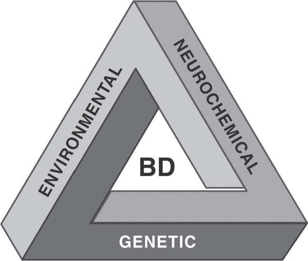
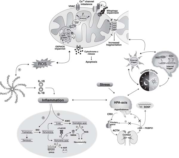
 PDF
 Print
 Send this article by email
 How to cite this article
 Submit a comment
 Mendeley
 Pocket
 Share on Facebook
 Share on Twitter