



ABSTRACT:
OBJECTIVE: To examine changes in plasma brain-derived neurotrophic factor (BDNF) levels in patients
undergoing electroconvulsive therapy or magnetic seizure therapy, as well as any early
or late variations in BDNF. Specific changes related to each technique were also evaluated.
METHODS: We measured plasma BDNF before and throughout treatment in adult patients (18 to 65
years old) diagnosed with treatment-resistant depression who underwent convulsive
therapy (electroconvulsive therapy or magnetic seizure therapy) in the ElectroMagnetic
Convulsive therapy for DEpression (EMCODE) project.
RESULTS: We enrolled 31 participants (mean age = 38.4 years, SD = 11.88), of whom 14 (45.16%)
underwent electroconvulsive therapy and 17 (54.84%) underwent magnetic seizure therapy.
Notable improvements in depressive symptoms were observed in both groups, with no
significant differences between them (p = 0.1046). However, no significant changes
in plasma BDNF levels were observed for either technique pre- or post-treatment (mean
difference = -93.01 pg/mL, 95%CI 545.88-359.86) or over time (coefficient = -67.95,
standard error = 37.75, p = 0.072).
CONCLUSIONS: The findings suggest that no significant changes occurred in plasma BDNF levels following
convulsive therapy, which challenges the notion that BDNF is a biomarker for treatment-resistant
depression.
Keywords: Treatment-resistant depression; electroconvulsive therapy; magnetic seizure therapy; brain-derived neurotrophic factor; neurophysiological biomarkers.
FIGURES
| Note: How to cite this article: Afonso-dos-Santos L, Cretaz E, Bellini H, Carneiro AM, Gallucci-Neto J, Tallib LL, et al. No changes in plasma brain-derived neurotrophic factor levels in treatment-resistant depression patients who underwent convulsive therapy. Braz J Psychiatry. 2025;47:e20243983. http://doi.org/10.47626/1516-4446-2024-3983 |
| Received: October 21 2024; Accepted: January 08 2025 |
INTRODUCTION
Depression, a prevalent mental health condition with a significant global impact, affects more than 264 million people worldwide.1 It is characterized by persistent sadness and a lack of interest or pleasure in previously rewarding or enjoyable activities. Up to 33% of patients who experience a depressive episode will develop treatment-resistant depression (TRD), defined as the lack of remission after at least two adequate first-line pharmacological trials regarding both duration and dosage.2,3 This highlights the need for both innovative treatment strategies and a deeper understanding of the mechanisms underlying TRD.3,4
Over the past 20 years, brain-derived neurotrophic factor (BDNF) has been studied for its role in the pathophysiology of depression and as an inducer of neuroplasticity. The interest in BDNF stems from the neurotrophic theory of depression, which proposes that deficits in neurotrophic support contribute to the development of depressive symptoms.5-7 Supporting this theory, previous clinical studies and meta-analyses have shown reduced blood levels of BDNF in patients diagnosed with depression compared to healthy controls, and they have also shown significant increases in BDNF levels after treatment with antidepressants.7-9
Electroconvulsive therapy (ECT) has long been recognized as an effective treatment for TRD.10,11 Magnetic seizure therapy (MST) has recently been introduced as a promising, less cognitively impairing alternative to ECT. Although it operates on a similar principle of inducing therapeutic seizures, MST combines characteristics of both ECT and repetitive transcranial magnetic stimulation. It generates electromagnetic stimulation through rapidly alternating magnetic fields, similar to repetitive transcranial magnetic stimulation, but in a more intensive manner, triggering a seizure once the individual's seizure threshold is exceeded. Like ECT, the procedure is minimally invasive and is conducted under general anesthesia with short-acting anesthetics and muscle relaxants. Magnetic fields are generated using a twin coil held at the vertex or on the prefrontal cortex.12,13
Prior systematic reviews have correlated ECT with increased blood levels of BDNF, underscoring its potential role in modulating neuroplasticity and alleviating depressive symptoms.14,15 However, more recent studies have contributed to a nuanced understanding of this relationship, suggesting that while ECT is associated with changes in BDNF levels, the strength of this correlation may be less pronounced than initially thought.16 This evolving evidence highlights the need for a more detailed examination of how ECT influences BDNF levels and the implications for its use in TRD. In addition, to our knowledge, no studies have evaluated BDNF in other convulsive therapies, such as MST.
In this study, we sought to examine possible changes in plasma BDNF levels in patients undergoing convulsive therapy. We also sought to observe early or late variations in BDNF, with assessments throughout treatment. Finally, we assessed specific changes related to the different techniques (MST and ECT), aiming to bring new insights to the field of convulsive therapies and TRD.
METHODS
Study design
This study is part of the ElectroMagnetic COnvulsive therapy for DEpression (EMCODE) project, including evaluations during two distinct phases of this project: the preliminary study, an open-label clinical trial employing MST in patients with TRD, and the ongoing randomized, controlled, double-blind clinical trial comparing the use of ECT and MST in patients with TRD. The preliminary study enrolled nine TRD patients undergoing MST treatment. These clinical results have already been published.17 The ongoing randomized clinical trial is comparing several clinical, cognitive, biological and neuroimaging outcomes in treatment-resistant patients who are being randomized to receive either ECT or MST.
Participant selection and recruitment
Individuals aged 18 to 65 years diagnosed with unipolar or bipolar TRD and experiencing a current depressive episode (baseline Hamilton Depression Rating Scale [HDRS-17] score > 16) were enrolled. All enrolled patients were eligible for ECT and provided informed consent. Diagnosis was confirmed by trained psychiatrists through the structured MINI interview.18
Patients were excluded from recruitment if they were pregnant, had other primary psychiatric disorders, presented depressive symptoms better explained by other clinical conditions, suffered from severe medical or neurological illnesses, had a history of psychoactive substance abuse in the last 6 months (excluding nicotine), recently used monoamine oxidase inhibitors, had neuromodulation treatment in the last year, were contraindicated for ECT or magnetic resonance imaging, or presented abnormalities in clinical laboratory parameters.
Interventions
In the pilot study, all patients received 12 sessions of MST twice a week. In the randomized clinical trial, patients were randomized to receive 12 sessions of ECT or 12-18 sessions of MST, also twice weekly. In the randomized clinical trial, up to 18 sessions were planned, with treatment concluding earlier if clinical remission was achieved by the 12th session, defined as an HDRS-17 score < 8.
The sessions were conducted in accordance with the ECT service protocols, following an adapted time-out (without informing the type or parameters of the procedure in cases where the patient was blinded to such information).
After monitoring, general anesthesia was administered by anesthetists experienced in ECT, with etomidate and succinylcholine for neuromuscular blockade. Atropine was also used to mitigate arrhythmia risks. The patients were monitored and received hyperventilation using a bag-valve-mask device with 100% oxygen. Bite blocks were used for further protection during stimulation sessions.
The ECT sessions were conducted using a Thymatron System IV device (Somatics LLC, Lake Bluff, IL, USA). Electrical parameters were set with a fixed pulse width of 0.5 ms and an amplitude of 0.9A. Electrodes were positioned bitemporally.10 The electrical charge for the initial session was titrated, starting at 5% of the charge and then doubling it until an effective seizure was induced or a maximum of three to four attempts were made. Once the convulsive threshold had been determined, the charge for the following sessions was 2.5 times this value.
MST sessions were administered using a MagVenture MagPro XP device with a TwinCoil positioned over the Fz point (MagVenture A/S, Farum, Denmark). The procedure mirrored that of an ECT session regarding anesthesia, oxygenation, and postictal recovery. Titration started with 100% amplitude at 100 Hz for 2 seconds, adding 2 seconds with each attempt until a seizure was induced or a maximum of three to four attempts were made. For the final charge in the following sessions, 4 seconds of stimulation were added, provided the total duration did not exceed 10 seconds.
Clinical evaluations
During the EMCODE trial, clinical and cognitive assessments were performed at baseline, after the sixth, twelfth, and eighteenth sessions, and again after 3 and 6 months. A variety of scales covering depressive symptoms, suicidal ideation, and neurocognitive evaluation was administered. Specifically, the HDRS-17, the Montgomery-Åsberg Depression Rating Scale (MADRS), the Young Mania Rating Scale, the Beck Depression Inventory (BDI), and the Clinical Global Impression were applied at every visit. For a detailed description of all assessments conducted in the EMCODE trial, see Bellini et al.19
For the main outcome, in alignment with previous BDNF and ECT research, this study compared pre- and post-treatment plasma BDNF levels. Pre-treatment levels were gauged at baseline, while post-treatment levels were assessed after the last therapy session, varying between the twelfth or eighteenth sessions based on patient progression.
As secondary outcomes, plasma BDNF levels were analyzed in various visits and clinical correlations were determined, examining the relationship between BDNF levels and depression scale scores.
Blood sample collection and brain-derived neurotrophic factor plasma analysis
Blood samples were collected at baseline, before sessions 6, 12, 18 (in patients who underwent up to 18 sessions), and at 4 weeks, 3 months, and 6 months post-treatment. All samples were collected in the morning, maintaining a minimum fasting period of 8 hours. The samples were analyzed using an enzyme-linked immunosorbent assay to quantify plasma BDNF levels.
Statistical analysis
The data were analyzed using Stata 18.0. Frequencies for categorical variables and statistical summaries for continuous variables were compared across groups using t-tests and chi-square tests. The t-tests were used to assess changes in plasma BDNF levels from baseline at each collection point and between treatment groups. Mixed-effect models were used to evaluate differences in BDNF levels at different collection times. Linear regression was applied to evaluate correlations between BDNF measures and clinical outcomes. A 5% significance level was used for all tests.
Ethics statement
Both study phases were approved by the internal institutional review board and the local and national ethics committees (decision 3.974.683, April 16, 2020). EMCODE is registered in Clinical Trials.gov (NCT05054699). For more details about EMCODE, see Bellini et al.19
RESULTS
Demographic and clinical characteristics of the sample
A total of 31 participants were enrolled whose mean age was 38.4 years (SD = 11.88, range 21-64). The sample was predominantly female, with 20 women (64.52%) and 11 men (35.48%). Most participants (26 individuals, 83.87%) were part of the clinical trial phase, while the pilot study included five participants (16.13%) (Table 1).
| Variable | n=31 |
|---|---|
| Sample | |
| Pilot study | 5 (16.13%) |
| RCT | 26 (83.87%) |
| Age (years)┼ | 38.4 (11.88), 21-64 |
| Sex | |
| Female | 20 (64.52%) |
| Male | 11 (35.48%) |
| Ethnicity | |
| Asian | 2 (6.45%) |
| Black/Mixed | 5 (16.13%) |
| White | 24 (77.42%) |
| Treatment modality | |
| ECT | 14 (45.16%) |
| MST | 17 (54.84%) |
| Diagnosis | |
| Unipolar | 22 (70.97%) |
| Bipolar | 9 (29.03%) |
| HDRS-17 score┼ | 23.39 (3.66), 18-35 |
| MADRS score┼ | 32.17 (4.60), 24-42 |
| BDNF plasma level on baseline (pg/mL)┼ | 1,552,097 (1,166,351), 137.86-4721.43 |
Data presented as n (%), unless otherwise specified.
BDNF = brain-derived neurotrophic factor; ECT = electroconvulsive therapy; HDRS-17 = Hamilton Depression Rating Scale; MADRS = Montgomery-Åsberg Depression Rating Scale; MST = magnetic seizure therapy; RCT = randomized clinical trial.
┼ Mean (SD), range.
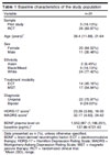
Regarding the type of depressive disorder, 22 participants (70.97%) were diagnosed with unipolar depression, and nine (29.03%) were diagnosed with bipolar disorder. The sample was primarily White (24 individuals, 77.42%), with five identifying as Black/Mixed (16.13%), and two identifying as Asian (6.45%).
The participants were divided between the treatment modalities, with 14 (45.16%) receiving ECT and 17 (54.84%) receiving MST. The groups were adequately homogeneous, with no significant clinical or sociodemographic differences at baseline. Both groups were predominantly unipolar. Although the MST group had a higher proportion of bipolar patients (n=7), the difference was non-significant (p = 0.101). For a more detailed description of the population data in the different treatment arms, see Supplementary Tables S1-S3.
In baseline clinical assessment, the mean HDRS-17 score was 23.39 (SD = 3.66, range 18-35), indicating moderate to severe depression. The mean MADRS score was 32.17 (SD = 4.60, range 24-42), which indicated significant depressive symptoms at the start of the study.
Change in pre- and post-treatment plasma brain-derived neurotrophic factor
There was no significant pre- and post-treatment differences in plasma BDNF levels (p = 0.68). The mean plasma BDNF level at baseline was 1539.21 pg/mL (SD = 1,211.96), while the post-treatment mean was 1,446.20 pg/mL (SD = 1128.18), with a mean difference (MD) of -93.01 pg/mL (95%CI 545.88-359.86) (Figure 1).

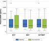
Our analyses revealed no significant differences BDNF level change between the ECT and MST groups. The ECT group had a non-significant mean increase of 16.87 pg/mL, while the MST group showed a non-significant mean decrease of 188.24 pg/mL. The calculated MD between the groups was 205.11 pg/mL, which was non-significant (95%CI -718.22-1128.43; p = 0.6517), indicating that the post-treatment variations in BDNF levels were comparable across both treatment types.
Change in plasma brain-derived neurotrophic factor during treatment and follow-up
In examining the time effects on plasma BDNF levels from baseline until the last follow-up at 6 months (Figure 2), mixed-effects model analysis revealed a non-significant decrease over time, with a time coefficient of -67.95 (standard error = 37.75, p = 0.072) (Figure 2).

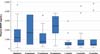
Upon analyzing the relationship between each specific visit and baseline plasma BDNF values, we found no significant difference at any endpoint, except for the 1-month post-treatment BDNF measurement. Paradoxically, there was a significant reduction in plasma BDNF levels at this time point compared to baseline in the paired t-test analysis, with a MD of -905 pg/mL (SD = 836.69) and a p-value of 0.002.
Separating the different treatment modalities, the results from the mixed-effects regression model indicated that for the ECT group, there was no significant change in plasma BDNF levels over time (coefficient = -8.38, p = 0.892), with substantial variability in individual baseline BDNF levels (variance = 1,208,345) and within-subject variability (variance = 699,379). Conversely, a significant decrease in BDNF levels was observed over time for the MST group (coefficient = -131.05, p = 0.003). The baseline variability among individuals (variance = 514,446) and within-subject variability (variance = 347,613) were also significant but were lower than in the ECT group. However, in an analysis of variance model, the effect in the MST group over the time was non-significant (p = 0.0839).
Plasma brain-derived neurotrophic factor and its correlations with depression measures
Regarding clinical depression data, paired t-tests showed significant reductions in depression scores across all scales. The MD in HDRS-17 scores decreased significantly (MD = -11.5667, p < 0.0001), indicating a notable improvement in depressive symptoms post-treatment. Similar significant improvements were observed in MADRS and BDI scores, with both showing a substantial post-treatment reduction (MADRS MD = -14.4444, p < 0.0001; BDI MD = -14.72, p < 0.0001).
No significant differences were observed between the ECT and MST groups for depression score change in any of the scales. The MD in HDRS-17 scores between the two groups was 4.53 (p = 0.1046); the MD in MADRS scores was 6.09 (p = 0.1185), while in BDI scores it was 9.08 (p = 0.0644). Although one group showed a consistent trend toward better outcomes, the difference was non-significant.
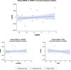
The baseline plasma BDNF levels did not predict changes in post-treatment HDRS-17 scores (p = 0.947). Regarding the impact of depression score variation on BDNF levels, linear regression using change in the depression scales correlated with the pre-post BDNF variation, showing no significant difference, with p = 0.400, p = 0.605, and p = 0.539 for the HDRS-17, MADRS, and BDI scales, respectively. These findings suggest that plasma BDNF levels do not correlate with the severity of depressive symptoms measured in these scales, as can be seen in Figure 3. There was no significant difference in plasma BDNF variation between bipolar and unipolar patients (p = 0.1682). In examining the influence of depressive episode duration on treatment outcomes, our regression analysis indicated no significant association between the length of the current episode and post-treatment changes in HDRS scores (ß = -0.0317, p = 0.343). There was no relationship between the duration of the current depressive episode and baseline BDNF level (ß = -5.297, p = 0.275), indicating that the chronicity of the current depressive episode did not significantly affect the magnitude of depressive symptom reduction or baseline BDNF levels in our sample.

DISCUSSION
The present study evaluated changes in plasma BDNF levels after ECT or MST in a sample of 31 TRD patients who were eligible for ECT. Contrary to our hypothesis, BDNF levels did not increase following a complete course of MST or ECT. Our results did not align with previous findings of a post-ECT increase in BDNF, since no significant changes were observed. The lack of a discernible change could be attributed to several factors, discussed below.
A notable aspect of our cohort was the presence of long-standing depression, with the participants having been treated with various psychopharmacological agents, which could have influenced baseline neurotrophic levels and the patients' response to convulsive therapy. This factor, coupled with substantial within-subject variability in BDNF levels, might have overshadowed any subtle changes induced by the treatment. It's essential to consider the high heterogeneity of BDNF findings reported in previous research, which underscores the complexity of using BDNF as a reliable state marker for depression. This contrasts with the suggestions of authors such as Piccinni et al.,20 who proposed that while plasma BDNF might act as a state-dependent marker, serum BDNF could be a trait marker for the disorder.
A subsequently proposed theory was that BDNF could increase relatively late after clinical improvement or the conclusion of treatment, peaking at up to 4 weeks.14 Nevertheless, this pattern was also not corroborated in our study, since 1 month after completing the sessions, plasma BDNF paradoxically decreased compared to baseline levels, challenging both the proposed late peak of BDNF and its utility as possible biomarker of disease state or treatment response in patients who undergo convulsive therapy.
In light of our findings, it is evident that the dynamics of plasma BDNF levels in the context of convulsive therapy for TRD are also not directly linked to clinical outcomes. The lack of significant change in post-treatment BDNF levels, in addition to the absence of a discernible relationship between BDNF level and depressive symptom severity, underscores the complexity of the neurobiological mechanisms underlying depression and its treatment. This complexity is further highlighted by the fact that baseline BDNF levels did not predict treatment response. Taken together, these findings suggest that plasma BDNF may not function as a direct biomarker for depression severity or treatment efficacy, and it may not play a significant role as a state marker of the disease, as proposed by Piccinni et al.20
These observations align with the nuanced perspectives described in prior reviews on the subject, which, despite noting an increase in blood BDNF following ECT treatment, could not establish a direct correlation between such increases and clinical improvement.14,16 Reinforcing the notion that plasma BDNF is not directly modulated by clinical depression outcomes, our study challenges the expectations set by a strict interpretation of the neurotrophic hypothesis of depression. This adds to the growing body of evidence suggesting that the relationship between peripheral BDNF levels and depression is complex and is mediated by multiple factors, warranting further investigation into the precise role of neurotrophic factors in depression and each treatment type.
The present study was pioneering assessment of BDNF in MST, providing a foundation for understanding BDNF dynamics in this relatively new treatment modality. However, the study's limitations, including its relatively small sample size and a high number of missing samples, particularly those not covering both pre- and post-treatment phases, should be acknowledged. These constraints, alongside the observed intra-individual variability, indicate a need for caution when interpreting our findings. Furthermore, we did not include baseline plasma BDNF measurements from a control group of healthy individuals. Although several studies have suggested a global trend toward reduced blood BDNF levels in patients with depression in comparison with the general population,7,9 it is not possible to confirm that such a trend specifically applied to our study population.
Future research, especially with larger cohorts and a randomized controlled design, is imperative to unravel the complexities of BDNF variability and evaluate its viability as a biomarker for treatment efficacy. Moreover, the neurotrophic hypothesis of depression advocates for a broader examination of neurotrophins and their corresponding receptors, including p75NTR, TrkB, and BDNF variants such as mature BDNF and its precursor molecule (pro-BDNF), to understand their intricate relationships and contributions to neurobiological mechanisms underlying treatment response.21-23 While earlier studies hypothesized that changes in plasma BDNF levels mirror adjustments in the central nervous system, recent inquiries have challenged this assumption, suggesting a more complex interplay.24,25 Consequently, future studies could greatly benefit from analyzing neurotrophic factors in cerebrospinal fluid, offering a more direct assessment of post-therapy changes in the central nervous system. This approach could provide crucial insight into the dynamics of neurotrophins like mature BDNF and pro-BDNF and their impact on depression treatment outcomes, further enriching our understanding of the neurotrophic hypothesis.
In conclusion, our investigation into changes in plasma BDNF levels in patients undergoing ECT and MST found no significant post-treatment changes. Although previous hypotheses have postulated plasma BDNF as a state marker for depressive disorders, our findings suggest that BDNF levels may not reflect immediate treatment-related changes, particularly in a cohort with TRD and a complex clinical history of medication use. This study, a pioneer in examining BDNF levels in MST, indicates the need for further research to understand the neurotrophic underpinnings of convulsive therapies. Given the above mentioned limitations, our results should be viewed with caution. Future research could benefit from exploring additional neurotrophins and their receptors, such as p75NTR and TrkB, and considering cerebrospinal fluid measurements of BDNF to provide a more direct assessment of central neurotrophic activity.
SUPPLEMENTARY MATERIALS
ACKNOWLEDGEMENTS
This project was partially funded by Fundação de Amparo à Pesquisa do Estado de São Paulo (FAPESP; process 2019/06009-6).
 PDF
 Print
 Send this article by email
 How to cite this article
 Submit a comment
 Mendeley
 Pocket
 Share on Facebook
 Share on Twitter