



ABSTRACT:
OBJECTIVE: White matter hyperintensities (WMH) are associated with major depressive episodes
(MDE) in individuals aged 65 and older. WMH are prevalent in adults under 65, yet
the association between their volume and MDE in this population remains uncertain.
This study aimed to assess the association of WMH volume with MDE and its severity
in patients aged < 65.
METHODS: Cross-sectional study (ancillary to clinical trial NCT02051413) of subjects under
the age of 65. Overall, 69 patients with MDE and 32 healthy controls (HCs) were included.
Severity was assessed with the Hamilton Rating Scale (HRS) and WMH were quantified
by two experts. Post-hoc mediation analyses were conducted if associations were found
between independent variables and WMH.
RESULTS: Mean age was 34.5 (12.4) years. There was no difference in WMH between patients and
HCs. Higher WMH volumes were observed in extremely severe MDE (2,170.2 [3,767.9] mm3 vs. 416.6 [594.9] mm3 [r = 0.21; p < 0.05]), which completely mediated the effect of age on severity.
CONCLUSIONS: In a sample of adults under 65, this study failed to identify higher WMH volume in
patients with MDE compared to HCs. However, WMH may act as a mediator of the association
between age and MDE severity. This finding suggests that WMH could contribute to more
severe depression in late life.
FIGURES
| Note: How to cite this article: Baudouin E, Corruble E, Gori P, Bloch I, Becquemont L, Duron E, et al. White matter hyperintensities and their role in major depressive episodes: a cross-sectional study in adults under 65. Braz J Psychiatry. 2025;47:e20243921. http://doi.org/10.47626/1516-4446-2024-3921 |
| Received: September 10 2024; Accepted: November 04 2024 |
INTRODUCTION
Major depressive disorder (MDD) is among the top ten contributors to health loss worldwide.1 Despite this, its pathophysiology and potential biomarkers remain unclear. This uncertainty notwithstanding, structural and functional brain changes, such as white matter lesions (WMLs), have been suggested to play a role in MDD and major depressive episodes (MDEs).2 WMLs are mainly caused by vascular pathologies, which can be categorized as small-vessel diseases and amyloid angiopathy. These abnormalities are best studied using magnetic resonance imaging (MRI), where they appear as white matter hyperintensities (WMH) on fluid-attenuated inversion recovery (FLAIR) sequences. WMH are highly prevalent in individuals aged 65 and older, with their prevalence increasing with age.3 Indeed, in a large cohort (1,077 subjects), WMH were found in 92% of participants.4
Furthermore, a correlation between overall WMH volume and MDE has been identified in a meta-analysis involving 8,498 patients,5 with an odds ratio (OR) and 95%CI of 1.37 (1.14-1.65). Specifically, deep WMH were found to be significantly associated with MDE (OR 1.47 [1.05-2.06]), whereas periventricular WMH were not. The same study identified a tendency towards a linear association between WMH volume and MDE severity. WMH significantly contribute to late-life depression (i.e., after age 65), often referred to as vascular depression.6 Vascular depression is hypothesized to be a major contributor to late-onset depression (i.e., that arising in late life in patients with no antecedent history of depression when younger) as opposed to early-onset depression (i.e., that present in late life in patients with a history of depression when younger).7 Definitions vary but there is a consensus on the importance of cerebrovascular pathology, with WMH playing a central role.8 Furthermore, there is a correlation between WMH volume and the severity of the late-onset depression.6
While the concept of vascular depression is typically applied to older adults, it is important to note the significant prevalence of WMH (5 to 50.9%9-11) in younger individuals as well. However, data on the relationship between WMH and MDE in subjects under the age of 65 are scarce12-15 and, to the best of our knowledge, no previous studies have identified a significant association. Therefore, there is a need for studies that quantify WMH volume, particularly in deep white matter tracts, to improve the understanding of the relationship between WMH and MDE in younger populations. Such insights could enable interventions to reduce the long-term burden of depression.
This study was designed as an exploratory analysis to investigate potential associations between WMH volume and MDE presence and severity in adults under 65. The secondary objectives were to assess the association between WMH volumes in deep white matter tracts and MDE presence and severity.
METHODS
Study design, setting, and participants
A cross-sectional ancillary study to the DEP-ARREST-CLIN study was conducted using inclusion data (ClinicalTrials.gov ID NCT02051413). This was a cohort study in which a total of 69 patients diagnosed with MDD experiencing MDE, who had been antidepressant-free for at least 1 month, were enrolled between February 2014 and January 2017. Additionally, 32 healthy controls (HCs) matched for age and sex were randomly selected from the telephone directory and included in the study. The inclusion criteria were as follows: a diagnosis of MDE (assessed with the Mini-International Neuropsychiatric Interview [MINI]16) and a 17-item Hamilton Depression Rating Scale (HDRS) score > 17 (maximum score 52 of 17 items, with higher score indicating more severe MDE17) in case of MDD.18 Patients with comorbid bipolar disorder, psychotic disorder, eating disorder, and/or addictions, according to DSM-5 criteria, were excluded. Patients were treated for 3 months with venlafaxine at a dosage left to their clinician's discretion. The neuroimaging and clinical data collected during this trial provided valuable resources for the investigation of WMH volume in relation to MDE severity in adults under 65 years old.
WMH volume assessment
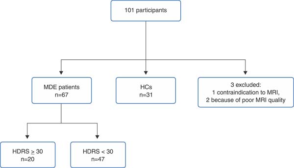
Brain structural MRIs (FLAIR sequences, 3T Philips Achieva) were performed at inclusion. One subject in the MDE group did not have an MRI because of an absolute contraindication (dental braces). Two further subjects, one from each group, were excluded from the study due to poor MRI quality. The specifications of the MRI scans are outlined in Supplementary Table S1.
WMH manual masks were created by two independent physicians, one of whom was an expert on WMH,19 who were blinded to group assignment. In the event of disagreement, the images were reassessed until complete agreement was reached. The images and masks were registered to the 1 mm3 Montreal Neurological Institute (MNI) space using the Advanced Normalization Tools (ANTs version 2.3.5)20 registration algorithm. We used the RegistrationSyNQuick routine to perform rigid and affine transformation using a 1-mm3 FLAIR template already registered to the MNI space21 as the fixed image. The total WMH volume was then calculated by summing the number of voxels, each of which had a volume of 1 mm3.
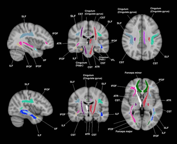
Next, the binary masks were registered to the Johns Hopkins University white-matter tractography atlas22,23 (Figure 1). The coordinates of the voxels were obtained using the FMRIB Software Library (FSL) v6.0.24-26 Regions were studied across both hemispheres by taking the mean of the left and right hemisphere regions with the number of voxels weighted in each hemisphere. This approach was adopted to avoid making any assumptions regarding any potential lateralized effect.27

The Fazekas rating scale,28 a commonly used clinical tool, was employed to assess the concurrent validity between the overall Fazekas global scores and the total WMH manual volumetric method. The Fazekas rating scale is scored on two subscales (periventricular WMH and deep WMH). Both range between 0 and 3, according to the size and confluence of the lesions, with higher scores indicating greater lesion burden. An overall estimation was calculated by summing the two subscales. Fazekas rating scale scoring was performed in a blinded manner, with each subject's group and volumetric assessment being unknown to the evaluators. All disagreements were reassessed by both evaluators until complete agreement was obtained. The weighted Cohen's coefficients of the initial rating step were calculated and are presented in Supplementary Table S2.
Depression severity
Depression severity was assessed using the HDRS-17 item total score, with scores of 24-29 indicating severe depression and scores of 30 or above indicating extremely severe depression.29-31
Statistical analysis
A Spearman correlation test was used to assess the correlation between WMH volume and Fazekas scores. Bivariate analyses were conducted on socio-medical data between MDE patients and HCs. This was done using the Wilcoxon rank-sum test for continuous variables and the chi-square test (or Fisher's exact test if size < 5) for categorical variables. A logistic regression was then conducted with MDE or HCs as the dependent variable. WMH volume, the primary outcome, was included in the model as an independent variable. The adjustment variables, chosen a priori, were age, sex, and a sum of cardiovascular risk factors (one or more among hypertension, ischemic heart disease, diabetes, dyslipidemia, and tobacco consumption). This score ranged from 0 to 5. The area under the curve (AUC) for different thresholds from the probabilities given by the model was calculated. In the event of a significant correlation between WMH volume and independent variables, a post-hoc mediation analysis was conducted in accordance with the Baron and Kenny method.32 This analysis consists of using several regression models to search for another variable that mediates the effect of an independent variable. Finally, subgroup analyses for MDE severity based on HDRS score were conducted using the same analysis plan with a linear regression model. The WMH volumes of the 10 deep tracts constituted the secondary outcomes.
The data were presented as mean (SD) for continuous variables and number (percentage) for categorical variables. As no missing values were identified for the variables of interest, no specific strategy was required to address missing data.
A power estimation indicated that 67 patients and 31 HCs would provide 80% statistical power with an expected mean difference (SD) of 600 (1,000) mm3 of WMH. A p-value of 0.05 was considered significant and Holm's procedure33 was applied to control the familywise error rate for deep white matter tracts analysis. All analyses were performed in R 4.1.2.34
Ethics statement
The authors assert that all procedures contributing to this work comply with the ethical standards of the relevant national and institutional committees on human experimentation and with the Helsinki Declaration of 1975, as revised in 2008. All participants provided written informed consent for study participation. This study was approved by the relevant ethics committee (Comité de Protection des Personnes Ile de France VI) and the French National Agency for Medicines and Health Products Safety (Agence Nationale de Sécurité du Médicament et des Produits de Santé [ANSM]).
RESULTS
Sample characteristics
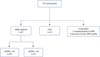
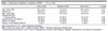
Ninety-eight participants were included in the analyses (Figure 2): 67 patients and 31 HCs. The mean age was 34.5 (12.4) years, 66 (67.3%) were female (Table 1), and 88 (89.8%) were right-handed.
| Total (n=98) | Patients (n=67) | HCs (n=31) | p-value | |
|---|---|---|---|---|
| Age, mean (SD) | 34.5 (12.4) | 34 (12.3) | 35.5 (12.9) | 0.57 |
| Female sex | 66 (67.3) | 45 (67.2) | 21 (67.7) | 0.96 |
| BMI, mean (SD) | 23.6 (4.8) | 23.6 (5.6) | 23.8 (2.6) | 0.21 |
| Educational level | ||||
| Post-secondary | 66 (67.3) | 44 (65.7) | 22 (71.0) | 0.60 |
| Cardiovascular risk factors┼ | 36 (36.7) | 27 (40.3) | 9 (29.0) | 0.28 |
| History of surgery | 43 (43.9) | 29 (43.3) | 14 (45.2) | 0.86 |
| History of neurological disease | 9 (9.2) | 7 (10.4) | 2 (6.5) | 0.72 |
Data presented as n (%), unless otherwise specified.
BMI = body mass index; HCs = healthy controls; HDRS = Hamilton Depression Rating Scale.
┼ Cardiovascular risk factors: one or more of hypertension, ischemic heart disease, diabetes, dyslipidemia, and tobacco consumption.
White matter hyperintensities volume
Concurrent validity of WMH volume with Fazekas score
Overall, Fazekas scores correlated significantly with WMH total volume assessed by manual masks (r = 0.94; p < 0.001).
WMH volume in MDE patients compared to HCs
WMH total volume was associated with age (r = 0.34; p < 0.001), but not with cardiovascular risk factors or sex (Supplementary Table S3).
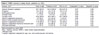
The overall mean WMH volume was 901.5 (1,992.8) mm3: 940.1 (2,233.2) mm3 in the patient group and 818.2 (1,363.1) mm3 in the HC group (p = 0.83). After adjustment by logistic regression for age, sex, and cardiovascular risk factors (AUC = 0.58), total WMH volume remained non-associated with MDE (p = 0.41). Regarding deep white matter tracts, no difference between groups was found (Table 2).
| Volume, mm3 | Total (n=98) | Patients (n=67) | HCs (n=31) | Unadjusted┼ p-value | Adjusted╬ p-value |
|---|---|---|---|---|---|
| Anterior thalamic radiation | 40.7 (93.5) | 44.3 (108.5) | 32.8 (47.3) | 0.87 | 0.39 |
| Cingulum | 4.2 (15.4) | 4.5 (17.0) | 3.6 (11.2) | 0.32 | 0.55 |
| Corticospinal tract | 5.1 (26.6) | 6.9 (32.0) | 1.3 (3.3) | 0.67 | 0.37 |
| Forceps major | 1.7 (7.3) | 1.6 (7.5) | 2.0 (7.2) | 0.28 | 0.90 |
| Forceps minor | 33.8 (83.8) | 38.4 (98.1) | 23.7 (36.9) | 0.60 | 0.29 |
| Inferior longitudinal fasciculus | 2.5 (9.2) | 3.2 (10.9) | 1.0 (3.2) | 0.95 | 0.18 |
| Inferior fronto-occipital fasciculus | 17.2 (56.3) | 20.9 (67.2) | 9.2 (15.7) | 0.77 | 0.27 |
| Superior longitudinal fasciculus | 17.8 (69.5) | 21.7 (80.5) | 9.3 (35.2) | 0.38 | 0.29 |
| Superior longitudinal fasciculus temporal part | 7.4 (30.8) | 9.5 (36.3) | 2.9 (11.3) | 0.32 | 0.31 |
| Uncinate fasciculus | 5.7 (18.1) | 7.3 (21.5) | 2.1 (4.8) | 0.97 | 0.20 |
WMH volume and MDE severity
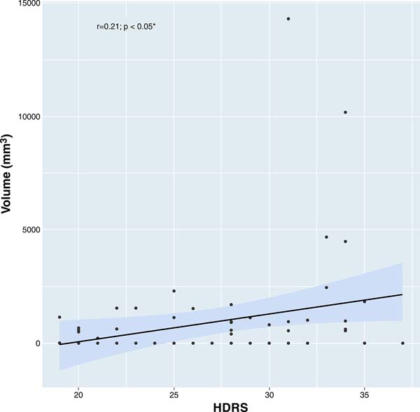
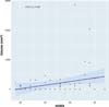
The mean (SD) HDRS score was 28 (5.3) out of 52. A non-significant trend was observed for an association between HDRS and age (r = 0.21; p = 0.09), but no association was found with sex, cardiovascular risk factors, or age at MDD onset (r = 0.06, p = 0.62; r = 0.01, p = 0.90; and r = 0.07, p = 0.54, respectively). A trend toward an association was observed between HDRS and WMH total volume (r = 0.21; p < 0.05) (Figure 3). However, this association did not remain significant after adjustment for age, sex, and cardiovascular risk factors (p = 0.10). WMH volumes in deep tracts were not associated with HDRS after Holm's procedure (Table 3).
| Volume, mm3 mean (SD) | Total n=67 | Spearman correlation's estimate | p-value | HDRS = 30 n=20 | HDRS < 30 n=47 | Wilcoxon's test p-value | Adjusted p-value┼ |
|---|---|---|---|---|---|---|---|
| Anterior thalamic radiation | 44.3 (108.5) | 0.21 | 0.09 | 93.1 (179.0) | 23.6 (46.9) | < 0.05 | 0.14 |
| Cingulum | 4.5 (17.0) | 0.12 | 0.33 | 11.2 (29.7) | 1.6 (4.7) | 0.10 | 0.34 |
| Corticospinal tract | 6.9 (32.0) | 0.27 | 0.03 | 18.1 (57.2) | 2.1 (6.4) | 0.09 | 0.38 |
| Forceps major | 1.6 (7.5) | 0.28 | 0.02 | 3.4 (12.8) | 0.8 (3.2) | 0.09 | 0.48 |
| Forceps minor | 38.4 (98.1) | 0.17 | 0.17 | 81.3 (161.4) | 20.2 (43.6) | 0.05 | 0.17 |
| Inferior longitudinal fasciculus | 3.2 (10.9) | 0.31 | < 0.05 | 6.5 (14.3) | 1.9 (8.9) | < 0.05 | 0.37 |
| Inferior fronto-occipital fasciculus | 20.9 (67.2) | 0.21 | 0.09 | 55.0 (116.6) | 6.3 (11.5) | < 0.05 | 0.12 |
| Superior longitudinal fasciculus | 21.7 (80.5) | 0.21 | 0.08 | 59.5 (140.5) | 5.6 (16.5) | < 0.05 | 0.14 |
| Superior longitudinal fasciculus, temporal part | 9.5 (36.3) | 0.22 | 0.08 | 26.8 (63.6) | 2.1 (5.7) | < 0.05 | 0.14 |
| Uncinate fasciculus | 7.3 (21.5) | 0.19 | 0.13 | 18.7 (36.5) | 2.5 (5.7) | < 0.05 | 0.09 |
HDRS = Hamilton Depressive Rating Scale; MDE = major depressive episode; WMH = white matter hyperintensities.
┼ Logistic regression adjusted for age, sex, and cardiovascular risk factors.
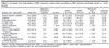
Severe MDE (HDRS = 24) vs. moderate MDE (HDRS < 24). Forty-four patients (65.7%) exhibited severe MDE (HDRS = 24). There was no significant difference in age, sex, or cardiovascular risk factors between patients with severe MDE and those with moderate MDE (1,227.6 [1,042] mm3 in the severe group vs. 390 [613] mm3 in the moderate group; p = 0.38). This result remained non-significant (p = 0.14) after adjustment for age, sex, and cardiovascular risk factors (AUC = 0.61). There was no difference in deep-tract WMH volumes between these subgroups (Supplementary Table S4).
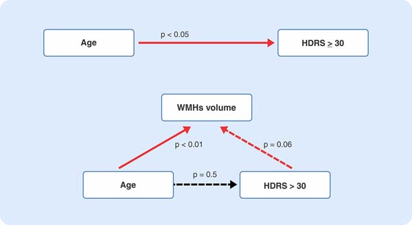
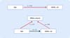
Extremely severe MDE (HDRS = 30) vs. mild/moderate/severe MDE. Twenty patients (29.9%) had an HDRS = 30. Age, sex, and cardiovascular risk factors did not differ significantly between MDE patients with HDRS = 30 and those with HDRS < 30. The extremely severe group had higher WMH total volume (2,170.2 [3,767.9] mm3 vs. 416.6 [594.9] mm3; p < 0.05), which did not survive adjustment (AUC = 0.66) for age, sex, and cardiovascular risk factors (p = 0.08). Mediation analysis demonstrated that the effect of age was mediated entirely by WMH volume (Figure 4).
The anterior thalamic radiation, superior longitudinal fasciculus, inferior longitudinal fasciculus, uncinate fasciculus, and inferior fronto-occipital fasciculus had a p < 0.05, but this did not survive Holm's procedure nor adjustment for age, sex, and cardiovascular risk factors (Table 3).
DISCUSSION
This study aimed to assess whether WMH volume was associated with MDE in adults under 65 years of age. We did not find a significant difference in WMH volume between MDE patients and HCs. However, the correlation between the severity of extremely severe MDE and age was mediated by WMH volume.
To the best of our knowledge, volumetric assessment of WMH has never been reported in MDE patients younger than 65, as in our study. The volume reported were in line with previous study on 428 HCs aged 65 and under,11 with a mean WMH volume of 1,386.42 (2,822.53) mm3 and a WMH prevalence of 50.9%. The absence of correlation between WMH volume and MDE in adults under 65 is also consistent with previous studies that employed semiquantitative ratings.12-14 This suggests that the relationship between WMH and depression may differ in younger adults. However, this does not exclude that subjects presenting with WMH at this age are not at risk of developing late-onset depression, as earlier onset implies high WMH loads.6 Further studies with other imaging modalities, such as functional imagery, may provide further insight on WML in MDE,27 as may prospective cohort designs.
Despite the absence of a significant difference in WMH volume between MDE patients and HCs, our findings indicated that MDE patients with more severe depression (HDRS = 30) exhibited larger WMH volumes, both overall and in specific deep white matter tracts. While this association did not remain significant after adjusting for age, sex, and cardiovascular risk factors, the results demonstrated a significant correlation between age and WMH volume. Given the established link between age and the severity of depression (e.g., higher risk of recurrence35), our findings suggest that WMH act as a mediator in the relationship between MDE severity and age. Therefore, it may be that subjects suffering from depression in late life, in which WMH are extensive,7 may have more severe illness in part because of these lesions.
This finding also raises questions regarding the nature of the relationship between depression severity and WMH. First, much as the severity of depression evolves over time, recent studies suggest that WMH could a potentially reversible pathological process, as they have been seen to regress36 with lower cognitive decline in certain populations. Moreover, treatments such as vasodilatation are being evaluated10 which supports the dynamic hypothesis.
Although greater progression of WMH volume is related to poorer outcomes in late-life depression,37 severe WMH does not necessarily lead to severe depression, as shown in people with specific white-matter diseases such as multiple sclerosis.38 Thus, although high WMH load may confer a higher risk for severe depression, this may be aggravated by pathological aging processes. Indeed, the concept of inflammaging - chronic low-grade inflammation occurring during pathological aging39 - is perhaps a lead, as depression has also a strong relationship with inflammation.6,40
Several limitations must be acknowledged. The primary limitation is the potential lack of statistical power to detect more nuanced differences in WMH volume and in specific deep white matter tracts between MDE patients and HCs. Although the DEP-ARREST-CLIN study had a robust design, it is important to note that it was not specifically designed to investigate WMH. However, our results are consistent with the current literature. Secondly, the semiquantitative rating method may be susceptible to inter-rater bias, although the initial Cohen's weighted coefficients were high (Supplementary Table S3). Thirdly, it is noticeable that WMH did not correlate with the cardiovascular score calculated in this study. Although this may be due to the calculation method, the low cardiovascular risk in a similar population41 may suggest that the WMH found in this study may not be from ischemic origins.11
In conclusion, in adults under the age of 65, this study failed to identify higher WMH volume in MDE patients compared to HCs. However, WMH may act as a mediator in the association between age and MDE severity. This finding suggests that WMH could contribute to more severe depression in late life. Future research on MDE severity should incorporate WMH volume data and age.
ACKNOWLEDGMENTS
This work was supported by a national grant from the Agence Nationale de la Recherche (ANR SAMENTA 2012) and sponsored by the Institut National de la Santé et de la Recherche Médicale (INSERM).
We gratefully acknowledge the contributions of the participants and the neuroradiology department.
 PDF
 Print
 Send this article by email
 How to cite this article
 Submit a comment
 Mendeley
 Pocket
 Share on Facebook
 Share on Twitter