



ABSTRACT:
The neurobiological factors associated with the emergence of major depressive disorder
(MDD) in adolescence are still unclear. Previous cross-sectional studies have documented
aberrant connectivity in resting-state functional magnetic resonance imaging (rs-fMRI)
networks. However, whether these findings precede MDD onset has not been established.
This scoping review mapped key methodological aspects and main findings of longitudinal
rs-fMRI studies of MDD in adolescence. Three sets of neuroimaging methods to analyze
rs-fMRI data were identified: seed-based analysis, independent component analysis,
and network-based approaches. Main findings involved aberrant connectivity within
and between the default mode network (DMN), the cognitive control network (CCN), and
the salience network (SN). Accordingly, we utilized Menon's (2011) triple-network
model for neuropsychiatric disorders to summarize key results. Adolescent MDD was
associated with hyperconnectivity within the SN and between DMN and SN, as well as
hypoconnectivity within the CCN. These findings suggested that dysfunctional connectivity
among the three main large-scale brain networks preceded MDD onset. However, there
was high heterogeneity in neuroimaging methods and sampling procedures, which may
limit comparisons between studies. Future studies should consider some level of harmonization
for clinical instruments and neuroimaging methods.
Keywords: Magnetic resonance imaging; major depressive disorder; longitudinal studies; functional neuroimaging; adolescent psychiatry.
FIGURES
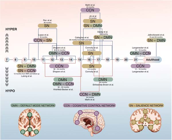
| Received: May 25 2021; Accepted: December 2 2021 |
INTRODUCTION
According to the Global Burden of Disease (GBD) study, major depressive disorder (MDD) is a leading cause of years lived with disability (YLDs).1 Its incidence starts to increase in early youth,2,3 and an adolescent-onset episode almost triples the risk of future episodes in adulthood,4 suggesting that neurodevelopmental factors affect the underlying pathophysiology of the disorder.5
In recent years, neuroimaging methods have enabled the exploration of neural mechanisms implicated in several psychiatric disorders.6 A particularly promising technique uses the functional magnetic resonance imaging (fMRI) signal called blood-oxygen-level-dependent (BOLD), which is a proxy for real-time brain activity in humans.7 At first, studies explored the fMRI BOLD signal while subjects performed tasks. Researchers then became interested in the baseline condition, before initiation of the task,8 known as resting-state fMRI (rs-fMRI). Since it carries fewer risks than other imaging modalities and fewer technical constraints, rs-fMRI is important for understanding the development of brain networks during adolescence and its relationship with the emergence of common psychiatric disorders, such as MDD.9
Recent reviews of rs-fMRI studies supported the hypothesis that MDD can be conceptualized as a brain network disorder.10,11 However, there are conflicting results, with studies reporting hyperconnectivity, hypoconnectivity, or even both. This is in contrast, for instance, with reward-task-based fMRI studies in MDD, which have consistently found less activation of reward circuitry regions, such as the ventral striatum, particularly in adolescence.12 In addition, the heterogeneity of methodological approaches to rs-fMRI data may impact direct comparisons between studies, limiting the ability to conduct adequate meta-analyses of neuroimaging.13-15 The neuroscience field, and neuroimaging studies in particular, has been struggling to perform well-powered investigations, with significant samples and methodological homogeneity.14 These issues have increased concerns regarding the reliability of findings from such studies to date. A systematic review and meta-analysis on this particular topic found that edges studied in fMRI research had an overall poor intraclass correlation coefficient.15 Although we focused on rs-fMRI, it is important to acknowledge that reliability problems seem even more prominent in task-based fMRI research. Vetter et al.,16 for instance, found that cognitive task findings were reliable, whereas emotional attention and intertemporal choice tasks had considerably better outcomes. Adding another challenge to the interpretation of neuroimaging findings, specific regions of interest (ROIs) have presented higher reliability, while others lacked consistency and varied considerably.17
The majority of brain network research on MDD has been performed in cross-sectional samples,10,11,18 which do not allow inferences on the temporality of the brain-behavior associations (i.e., which came first). It is also uncertain whether adult findings apply to adolescence, when biological aspects of neurodevelopment are still taking place.19 In addition, relevant differences in clinical MDD profiles exist between adults and adolescents.20 Methodological aspects may also differ significantly between these populations, such as diagnostic criteria, clinical interviews, and sampling criteria. Even though previous reviews investigated altered brain networks in MDD, they were not focused on adolescent MDD,10,11 longitudinal studies,18 or rs-fMRI.21
This scoping review maps the literature addressing the research question: what is the evidence from longitudinal studies linking altered rs-fMRI brain networks to adolescent MDD? Specifically, our aims were dual: first, to map key methodological elements of rs-fMRI research and how they were employed in adolescent MDD studies; second, to investigate if there is evidence from longitudinal studies suggesting that aberrant resting-state connectivity precedes MDD.
METHODS
We followed systematic procedures suggested by the Preferred Reporting Items for Systematic reviews and Meta-Analyses extension for Scoping Reviews (PRISMA-ScR).22 As required by the PRISMA-ScR protocol, study and/or personal fundings for included studies/authors were collected and are listed in Table S1, available as online-only supplementary material. The review protocol was not registered. The eligibility criteria were studies reporting on: 1) adolescence (10-20 years of age); 2) depression (categorical MDD or dimensional measures); 3) rs-fMRI; iv) longitudinal designs. The exclusion criteria were: 1) interventional designs; and 2) task-based functional connectivity. We performed an online search using the PubMed/MEDLINE database in August 2020 using the keywords (Adolesc*) AND (Depress*) AND (rest*) AND (connect*). Titles and abstracts of the search results were independently screened by two authors (MM and PP) using the online platform Rayyan.23 Data from included studies was charted by one author (MM) and then independently revised by a second author (PP). Core variables for extraction were defined on the basis of previous reviews in the field.18,21 References from relevant reviews and commentaries were also screened.
To provide a critical appraisal of the evidence, we used a rationale to synthesize data according to the triple-network model.6 In a seminal paper, Menon 6 critically reviewed the fMRI literature and proposed a triple-network model for neuropsychiatric disorders. According to this model, dysfunctional connectivity between or within the three most replicated brain networks was associated with emotional and behavioral symptoms. These networks are the default mode network (DMN), the cognitive control network (CCN) (also called the central executive network [CEN]), and the salience network (SN).6,24-28 Finally, we categorized our findings according to the specific network connectivity pattern: hyperconnectivity, hypoconnectivity, or mixed. We describe in detail, in the Results section, the assumptions or simplifications that were necessary to adequately classify main study findings according to this proposal, such as assigning a specific region to a network in a seed-based analysis study.
RESULTS
Aim 1 - Key methodological elements of resting-state functional magnetic resonance imaging (rs-fMRI) research
The correlation of the BOLD signal time-series at rest exhibits consistent patterns of synchronous activity among different brain regions.29 This pattern of co-activation or co-deactivation is called intrinsic functional connectivity (iFC).9
Since there is no task to perform in rs-fMRI, the researcher asks the patient to stare at a cross mark during the entire scan. However, studies showed that even across distinct resting-state conditions, such as sleep and anesthesia, these iFC patterns were highly consistent.29
The duration of the scan protocol varies from a few minutes to half an hour, and several methods now allow researchers to remove undesirable artifacts of the BOLD signal, such as the effect of minimal head movements during the scan.8,30,31
There are three main sets of methods to analyze brain networks using rs-fMRI:
- Seed-based analysis: investigates whether the BOLD signal time-series of a predetermined ROI (i.e., the seed) is correlated to any other brain region (seed-to-whole-brain) or to another predetermined ROI (seed-to-seed). Seeds can be defined using data from previous studies or by applying hypothesis-driven theoretical assumptions.32
- Independent component analysis (ICA): a hypothesis-free methodology in which the correlations of the BOLD signal time-series (i.e., connectivity) are explored for the whole brain.33 Therefore, there is no need for a predetermined ROI selection. Researchers then interpret inter-regional correlation patterns captured by ICA to ascertain for existing brain networks.
- Network-based approach: this branch encompasses various methods derived from graph theory analysis, the mathematical study of networks.34 It investigates which nodes (predetermined ROIs) and edges (ROI-ROI BOLD time-series correlations) are relevant in the context of a specific network. Several measures of network features (i.e., integration or segregation) and performance (i.e., nodes centrality, local, and global efficiency) can be explored.
ICA-based rs-fMRI studies revealed that the spontaneous activity of the brain can be organized in distinct networks.35 Several networks have been reported, such as auditory, basal ganglia, primary and secondary visual cortices, language, and sensorimotor networks. Specific connectivity patterns can be interpreted as a functional specialization. However, according to the triple-network model, the most significant networks for neuropsychiatric disorders are the DMN, the CCN, and the SN.6
Default mode network (DMN)
This network consists of brain regions that are synchronously activated during periods without any specific task assigned.25,36 The DMN is divided into anterior and posterior sub-networks. The main node of the anterior DMN is the medial prefrontal cortex (mPFC). This node plays a particular role in emotional regulation and self-referential processes, showing important connections with subcortical regions, such as the amygdala.37,38 The posterior DMN encompasses the posterior cingulate cortex (PCC) and the precuneus cortex. These nodes are involved in consciousness and memory processing, displaying relevant connections with the hippocampal formation.39-41 Other brain regions are also part of the DMN, such as the inferior parietal cortex, the lateral temporal cortex, and the subgenual anterior cingulate cortex (sgACC).40,42,43
Cognitive control network (CCN)
The CCN is also known as the CEN or the "task-positive network". In contrast to the DMN, the CCN is highly activated during cognitive tasks,44 although the presence of the CCN in rs-fMRI data is a highly replicable finding.32 The CCN and the DMN are commonly referred to as "opposite networks".45,46 The CCN consists mainly of frontoparietal regions such as the dorsolateral prefrontal cortex (dlPFC), the posterior parietal cortex, and the dorsal anterior cingulate cortex (dACC). It has been implicated in regulatory top-down control and in specific cognitive functions, such as decision-making and working memory.47,48
Salience network (SN)
The main regions of the SN are the fronto-insular cortex, the amygdala, and the ventrolateral PFC (vlPFC). Altogether, these regions have been implicated in tasks involving emotionally relevant stimuli.26 SN is also relevant to the detection of environmental cues by mediating the constant alternation between the DMN and CCN activation.26,49,50
Resting-state fMRI and neurodevelopment
rs-fMRI is particularly useful for studying neurodevelopment in children and adolescents.31 Understanding complex task instructions, for instance, may be difficult in childhood. Consequently, numerous studies used this method to leverage knowledge on typical neurodevelopment, showing segregation and specialization of large-scale networks across development.31,51,52 The centrality of the subcortical and cerebellar nodes among a whole-brain network, for instance, decreases from late childhood to early adolescence.53 In contrasting, the relevance of cortical nodes progressively increases in the same developmental window. These findings are in line with structural changes in cortical regions across development,54 supporting the hypothesis of late maturation for cortical regions - particularly the PFC.55,56 In sum, important neurodevelopmental changes occur in the adolescent brain,27,56,57 which may impact rs-fMRI findings as they relate to MDD.
Aim 2 - Evidence suggesting that aberrant brain connectivity precedes adolescent-MDD onset
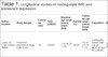
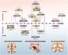
Our literature search retrieved 307 research articles. Eighteen studies met inclusion criteria and were retrieved for full-text analysis. Five studies were subsequently excluded due to interventional design (n=3, cognitive-behavioral therapy; n=1, transcranial magnetic stimulation; n=1, antidepressant). The 13 remaining articles were included in this review. Table 1 summarizes the main findings of these studies. Figure 1 depicts the developmental periods explored in each study and whether findings point to hyperconnectivity, hypoconnectivity, or to a mixed pattern.
| Author, location | Study sample | Follow-up (mo)┼ | Total (n/MDD) | Baseline age range (years) (mean [SD]) | Female (%) | Follow-up age range (years) (mean [SD]) | Population | Method | fMRI scans | MRI model/scan duration | Depression assessment | Main analyses | Secondary analyses | Findings | Specificity analyses | Network model interpretation | Limitation |
|---|---|---|---|---|---|---|---|---|---|---|---|---|---|---|---|---|---|
| Lopez,58 St. Louis, United States | Preschool Depression Study (Luby et al.59) | 18 | 143/58 | 7-12 (9.74-1.23) | 66.00 | 10-16 (12.52-1.11) | Community-based, high-risk,58 MDD-hx, and 85 controls | Seed to whole brain | 2╬ | 3-T Tim Trio/6.8 min | Child and Adolescent Psychiatric Assessment (categorical); CDI and Child Sadness Management Scores (dimensional) | dlPFC and amygdala | vlPFC, insula, vmPFC, dorsal anterior cingulate cortex | Amygdala, striatum and PFC network (within-circuit) linked to concurrent and future MDD | Other diagnoses, anxiety, disruptive | Hyperconnectivity, CCN-SN | No categorical MDD analysis; no significant finding after adjusting for concurrent depressive symptoms |
| Pan,60 São Paulo and Porto Alegre, Brazil | BHRCS | 36 | 637/56 | 6-12 | 45.6 | 9-15 (10.6-1.9) | Community-based, high-risk | Graph theory analyses | 1 | 1.5-T GE Signa HDX/6 min | Developmental and Well-Being Assessment | Node strength (i.e., degree centrality) of striatal nodes within a 11-node reward network | Node strength (i.e., degree centrality) of other nodes: thalamus, insula, pre-SMA, ventral tegmental area, anterior cingulate cortex, PCC, vmPFC | Increased left ventral striatum connectivity within reward network predicted a 50% increase in MDD risk at follow-up | Attention-deficit/hyperactivity disorder, Anxiety, any substance use | Hyperconnectivity, within reward network | Investigated only iFC within one brain network, no cross-network analyses |
| Davey,61 Melbourne, Australia | ADS | 24 | 56/8 | (16.5-0.5) | 44.60 | 19 (18.8-0.5) | Community-based, high-risk, eight new-onset MDD at follow-up | Seed to seed | 2 | 3-T Siemens MAGNETOM Trio/not reported | CES-D | Amygdala to sgACC associations with negative affectivity | Longitudinal associations with MDD onset | Concurrent increase in amygdala -sgACC connectivity among new-onset MDD participants; amygdala -sgACC connectivity changes associated with negative affectivity changes | Attention-deficit/hyperactivity disorder, anxiety, any substance use | Hyperconnectivity, SN-DMN | MDD was a secondary analysis |
| Luking,62 St Louis, United States | Early Emotional Development Program at Washington University School of Medicine in St. Louis | 48-60 | 51/26 | 3-6 | 52.90 | 7-11 | Clinical sample; four subgroups: MDD-hx, familial high-risk, both combined, healthy controls | Seed to seed | 2╬ | 3-T Tim Trio Scanner/6.8 min | Preschool Age Psychiatric Assessment Categorical (previous); CDI, dimensional (current) | Amygdala to bilateral: superior temporal gyrus, hippocampus; right: putamen and para hippocampal gyrus; left: inferior temporal gyrus; middle frontal gyrus, superior middle gyrus, IPL, precuneus, superior frontal gyrus | Associations with CDI and MDD severity | Reduced connectivity between amygdala and the dlPFC, dmPFC, cingulate cortex, hippocampus, and hippocampal gyrus in MDD/family risk | Other diagnoses (not specified) included in control group and used as covariates | Hypoconnectivity, SN-DMN, SN-CCN | MDD episode occurred several years before the fMRI |
| Langenecker,63 Ann Arbor and Chicago, United States | Convenience sample from University of Illinois | 13-18 | 109/60 (21 recurrent MDD) | 18-23 (21.00-1.38 for recurrence groups) | 54.10 | 19-25 | Community-based convenience, high-risk, 60 with MDD-hx, 21 MDD recurrence at follow-up | Seed to whole brain | 1 | 3-T, GE Scanner/8 min | Diagnostic Interview for Genetic Studies | sgACC and middle frontal gyrus | rs-fMRI analyses are part of a larger task-based study | Increased connectivity between sgACC, middle frontal gyrus, and other CCN regions, and decreased connectivity between the middle frontal gyrus and parietal regions in MDD-Hx who presented an MDD recurrence at follow-up | Test-retest intraclass correlation coefficient analyses with fMRI scans after 4-12 weeks | Hyperconnectivity, DMN-CCN; hypoconnectivity within CCN | Previous history of MDD defined retrospectively |
| Callaghan,64 Melbourne, Australia | ADS | 36 | 101/14 | 16 (16.5-0.53) | 46.50 | 19 (18.8-0.5) | Community-based, 14 developed MDD prior to fMRI scan (excluded from analyses) and 14 after | Seed to whole brain | 1 | 3-T Siemens MAGNETOM Trio/not reported | K-SADS-E | Amygdala to whole brain. Associations between previous maternal aggression and current amygdala iFC. | VS and Nac with whole brain | Increased amygdala-temporal cortex and amygdala-insula iFC in MDD group. Findings mediated the maternal aggression-MDD association. | VS and Nac iFC was not associated with maternal behavior | Hyperconnectivity, within SN | Relatively small sample size for MDD group |
| Hirshfeld-Becker,65 Boston, United States | Convenience sample from Massachusetts General Hospital | 38,7-60,2 (47.8, 4.5) | 41/10 | 8-14 (11.0-1.72) | 46.30 | 12-18 (15.3-1.7) | Community-based, family high-risk MDD, 10 at risk developed MDD | Seed to whole brain | 1 | 3-T TrioTim Siemens Scanner/6.2 min | K-SADS-E and CDI | mPFC, PCC, SgACC, dlPFC and amygdala | Exploratory seed-to-seed amygdala-dlPFC | Incident MDD group exhibited weaker connection between sgACC and IPL and between left and right dlPFC. Non-converters (resilient to the genetic risk) showed higher sgACC-IPL iFC connectivity. | Support vector machine classifier using iFC | Hypoconnectivity, within DMN and between DMN-CCN | Relatively high attrition rate (28%) and few cases of incident MDD |
| Jin,66 New York, United States | ADEPT | 18 | 173/none | 13-15.5 (15.29-0.65) | 100.00 | 14.5-17 | Community-based, convenience | Graph theory analyses | 1 | 3-T TrioTim Siemens Scanner/5-6 min | IDAS-II | A within-circuit graph including PFC, amygdala and striatum and the iFC between this circuit with the whole brain (extended-circuit) | A whole-brain graph using 217 nodes previously defined in a rs-fMRI atlas | Amygdala, striatum, and PFC network (within-circuit) linked to concurrent and future MDD | Extended-circuit model did not increased sensibility in predicting MDD symptoms | Hyperconnectivity within SN and between SN-DMN§ | Included only female participants |
| Strikwerda-Brown,67 Melbourne, Australia | ADS | 24 | 72/11 | (16.47-0.59) | 45.80 | (18.75-0.48) | Community-based, 72 adolescents, 11 MDD on sets between follow-ups 2 and 6 with high scores at each time point | Seed to whole brain | 2 | 3-T Siemens MAGNETOM Trio/12 min | CES-D | Longitudinal analyses of sgACC iFC | Cross-sectional analyses of sgACC iFC | Decreased iFC between sgACC and dmPFC, PCC, right angular gyrus, and left middle temporal gyrus associated with higher depressive symptoms at follow-up | Models adjusted for anxiety symptoms | Hypoconnectivity within DMN | Longitudinal findings did not survive to head movement adjustment for both time points |
| Shapero,68 Boston, United States | Convenience sample from Massachusetts General Hospital of Illinois | 47.4 (38.1-54.7, 4.68)|| | 44/12 between scans; two at follow-up | 8-14 (11.0-1.72) | 45.00 | 11-19 (14.3-1.9) | Community-based, genetic high-risk MDD, 11/28 high-risk and 1/16 low-risk developed MDD | Seed to whole brain | 1 | 3T TrioTim Siemens Scanner/6.2 min | K-SADS-E both times; CDI and Child Behavior Checklist at follow-up | DMN seeds (mPFC, PCC), CCN seeds (bilateral dlPFC) and amygdala | Increased functional connectivity between DMN and supramarginal gyrus and decreased connectivity within the CCN (between left and right dlPFC) predicted onset of MDD | Self- ad parent-reported dimensional symptomatic change | Hyperconnectivity DMN-CCN; hypoconnectivity within CCN | Canonical correlation analyses to reduce rs-fMRI variables due to low sample size | |
| Connolly,69 San Diego, United States | Convenience sample recruited from adolescent psychiatric and primary care clinics | 3.3 (0.60)|| | 101/48 MDD (24 at follow-up) | 13-18 (16.1-1.3) | 61.30 | Only 3 months of follow-up | Clinical sample, 48 drug naive MDD adolescents, 24 completed follow-up | Seed to whole brain | 1 | 3T GE MR750/8.32 min | Children's Depression Rating Scale-Revised | Amygdala to whole brain | Increased connectivity from R. Amygdala to orbital middle frontal gyrus was associated with greater symptoms at follow-up; increased connectivity from R. Amygdala to bilateral insulae was associated with a reduction in follow-up depressive symptoms. | Hypoconnectivity and hyperconnectivity within SN | High follow-up attrition rate; treatment type was not controlled | ||
| Malhi,70 Sydney, Australia | Convenience sample recruited from the same school | 24 | 88/27 symptomatic | 14-16 (15.35-0.52) | 100.00 | 16-18 | Community-based from a single school, follow-up n=71 (27 with emotional symptoms and 44 controls) | Independent component analyses | 1 (dynamic fMRI) | 3-T Siemens MAGNETOM Trio Scanner/not reported | Child and Adolescent PsychProfiler, CDI | 28 components/networks comprising DMN (n=7), CCN (n=7), and attentional networks (n=14) | Longitudinal changes in CDI and state anxiety | Left lPFC showed weaker connection with posterior and anterior midline PFC (DMN) and greater connection with right lPFC (CCN) in symptomatic girls | Increased left LPFN-right LPFN iFC contributed to longitudinal CDI changes | Hypoconnectivity CCN-DMN; hyperconnectivity within CCN | Sample with low representativeness; only girls assessed; no formal MDD diagnosis |
| Jalbrzikowski,71 Pittsburgh, United States | Accelerated cohort longitudinal design study Replication used the Philadelphia Neurodevelopmental Cohort | 15-45 | 246/none (anxiety and depressive symptoms) | 10-22 longitudinal sub-sample; 20-25 cross-sectional subsample | 49.10 | Not reported | Community-based, participants and their first-degree relatives without psychiatric disorder. Follow-up data for two (n=117) or three (n=90) visits. | Seed to seed | 1-3 | 3-T Tim Trio/5 min | Youth Self-Report, 10-17 years-old/ Adult Self-Report, ages 18-25 years-old | Amygdala (BL and CM subregions) and vmPFC (anterior vmPFC, anterior and posterior OFC, rACC, ventral anterior cingulate cortex, sgACC subregions) | - | Increased centromedial amygdala-rACC connectivity was associated with anxiety and depression symptoms in early adulthood | Time-varying effect models evaluated differential effects of age, sex, and emotional symptoms | Hyperconnectivity SN-DMN | No formal MDD diagnosis |
ADS = Orygen Adolescent Development Study; BHRCS = Brazilian High-Risk Cohort Study; CCN = cognitive control network; CDI = Child Depression Inventory; CES-D = Center for Epidemiological Studies Depression Scale; dlPFC = dorsolateral prefrontal cortex; DMN = default mode network; dmPFC = dorsomedial prefrontal cortex; fMRI = functional magnetic resonance imaging; iFC = intrinsic functional connectivity; IPL = inferior parietal lobule; ITG = inferior temporal gyrus; K-SADS-E = Kiddie Schedule for Affective Disorders and Schizophrenia for School-Aged Children - Present and Lifetime Version; lPFC = lateral prefrontal cortex; MDD = major depressive disorder; MDD-hx = major depressive disorder history; mPFC = medial prefrontal cortex; Nac = nucleus accumbens; PCC = posterior cingulate cortex; PFC = prefrontal cortex; rACC = rostral anterior cingulate cortex; rs-fMRI = resting-state functional magnet resonance image; SD = standard deviation; sgACC = subgenual anterior cingulate cortex; SN = Salience Network; vmPFC = ventromedial prefrontal cortex; VS = ventral striatum.
┼ Original values in weeks. Calculated in months by the author for standardization.
╬ Both scans performed at same time point.
§ See main text for limitations of this interpretation.
|| Mean (standard deviation).
The first rs-fMRI longitudinal study in adolescent MDD was published in 2011.62 Low- and middle-income countries (LMIC) were underrepresented, with only one study from Brazil60 as compared to eight from the United States58,62,63,65,66-69,71 and four from Australia.61,64,67,70 Sample sizes ranged from 41 to 637 subjects.60,65 Smaller samples reported on well-characterized clinical MDD,63-65,69 whereas larger studies were frequently drawn from community-based samples in which categorical MDD assessment was not commonly performed.58,61,62,66-68,70,71 Some of these community-based studies only reported dimensional measures of depressive symptoms from specific (Child Depression Inventory, Center for Epidemiological Studies Depression scale) or non-specific (Youth Self-Report and Adult Self-Report, Child and Adolescent PsychProfiler) instruments.
Several high-risk strategies were adopted to select participants. These can be categorized into: 1) family history of MDD or other non-specific family psychiatric morbidity;60,65,68 2) previous individual history of MDD;58,63,69 and 3) individual high risk due to phenotypic or temperamental traits.61,70 One study used a mixture of these strategies.62 Individual psychiatric comorbidity was allowed in some studies for both case (MDD) and control groups.58,60 The amygdala was the most studied ROI,58,61,62,64,65,68,69,71 while the most frequent methodological approach was seed-based analysis.58,61-65,67-69,71 Only three studies employed repeated fMRI scan assessments.61,67,71
The following sections summarize main findings from each study according to the analytical rs-fMRI approach and the directionality of the brain-behavior association, vis-à-vis Menon's triple-network model.6 Overall, four studies reported on mixed findings of hyperconnectivity and hypoconnectivity, six studies reported only hyperconnectivity, and three reported only hypoconnectivity among the three networks (DMN, CCN, and SN).
Hyperconnectivity findings for seed-based analysis studies
Using a seed-to-seed approach, a study found that higher negative affectivity was associated with increased connectivity between the amygdala and the sgACC over 24 months of follow-up.61 Davey et al.61 measured negative affectivity (NA), a temperamental trait previously associated with increased risk for future MDD, with the revised Early Adolescent Temperament Questionnaire. They defined high-risk participants based on increased levels of NA. Interestingly, among the 56 high-risk adolescents, new-onset MDD was associated with follow-up concurrent increased amygdala-sgACC connectivity. These results suggest a pattern of hyperconnectivity between the SN and the DMN.
Another study from the same group linked exposure to observed maternal aggressive behavior in early adolescence (at 12 years old), abnormal amygdala iFC in mid-adolescence, and MDD at 19 years old.64 Callaghan et al.64 followed 101 children for over 7 years (3 years between fMRI scan and MDD assessment) and found increased connectivity from the amygdala to temporal cortices and bilateral insula in MDD. It is noteworthy that this finding mediated the association between maternal aggressive behavior and MDD. In a specificity analysis, striatal seeds did not show the same pattern of association, indicating a specific role for the amygdala in the relationship between early trauma and adolescent-onset depression. We categorized these findings as evidence of SN within-network hyperconnectivity.
Using a high-risk design based on previous depressive symptoms, the study from Lopez et al.58 included 58 adolescents with a history of MDD (MDD-hx) and 85 adolescents without previous MDD episodes (no-MDD group). Data were drawn from a large 12-year longitudinal study, the Preschool Depression Study. Although baseline recruitment study assessed familial risk (for more details, see Luby et al.59), Lopez et al.58 considered personal history of MDD as the only high-risk criteria. Importantly, the no-MDD group could also include subjects with other previous or ongoing psychiatry disorders such as attention-deficit/hyperactivity disorder, anxiety, and conduct disorder. Results suggested that hyperconnectivity between the dlPFC and the dorsal ACC in preadolescence (9-14 years old) predicted higher levels of depressive symptoms in adolescence (10-16 years old). These results were not statistically significant when models included baseline depressive symptoms concurrent to the brain scan. Additionally, there were no categorical MDD assessments at follow-up. Keeping in line with Menon's triple-network model, these findings suggest a putative increased connectivity between the CCN and the SN, which comprises dlPFC and dACC regions, respectively.
Jalbrzikowski et al.71 was the only study that included up to three rs-fMRI scans from the same subject. They assessed connectivity between the amygdala and the ventromedial prefrontal cortex (vmPFC) using a seed-to-seed approach in a large sample of adolescents and young adults. The focus of the study was to establish normative patterns of functional and structural (white-matter tract) connectivity between the amygdala and the vmPFC across youth. A normative pattern of decreasing amygdala-vmPFC connectivity between the ages of 10-25 years was established and then replicated in an independent sample. Increased amygdala-ACC connectivity was linked to higher levels of internalizing psychopathology - a mixture of depression and anxiety symptoms - in late adolescence and early adulthood. We categorized these findings according to the seed regions as putative evidence of SN-DMN hyperconnectivity.
Hypoconnectivity findings for seed-based analysis studies
Luking et al.62 used a seed-to-seed approach to investigate amygdala connectivity in a high-risk sample. They employed a mixture of individual and family high-risk factors to compose four non-overlapping groups: MDD-hx, familial high-risk, both combined, and none of these conditions (healthy controls). Repeated standardized clinical assessments were used to ascertain for previous MDD episodes.62 This strategy resulted in a low number of participants for each group. Nevertheless, they were able to follow participants for up to 5 years. MDD-hx and familial high-risk groups exhibited decreased connectivity between the amygdala and several brain regions, including the dlPFC, the dmPFC, and the hippocampus. Intriguingly, the same pattern of aberrant connectivity was not found in the combined MDD-hx plus genetic risk group. Since this study investigated MDD several years before the rs-fMRI scan, it was not possible to disentangle whether connectivity alterations represent high-risk patterns or adaptations to previous MDD episodes. We categorized these results as between-network hypoconnectivity from the SN to the DMN and the CCN.
Hirshfeld-Becker et al.65 selected a high-risk sample based on family history of MDD, assessed at baseline with semi-structured diagnostic instruments.65 Parental past or current episodes of mood disorders (MDD, bipolar disorder, or dysthymia) were considered a high-risk factor for probands. Results indicated decreased connectivity between left and right dlPFC among adolescents who developed MDD at follow-up. This group exhibited weaker connectivity between the sgACC and the inferior parietal lobule (IPL), whereas "non-converters" (i.e., those resilient to the genetic risk) showed increased sgACC-IPL connectivity. Therefore, we found evidence of within-network hypoconnectivity for CCN and DMN-CCN between-network hypoconnectivity.
Using sgACC seeds and a seed-to-whole-brain approach, Strikwerda-Brown et al.67 found an association between decreased sgACC-dmPFC connectivity and incident depressive symptoms. Concurrent and longitudinal associations with depressive symptoms were also observed with other posterior DMN nodes, such as the PCC, suggesting a pattern of hypoconnectivity within the DMN. Longitudinal associations, however, did not survive when head motion was included as covariate for both time points. Few subjects developed full-blown MDD episodes at follow-up, which may have limited the statistical power to perform categorical MDD analysis.
Mixed findings for seed-based analysis studies
A longitudinal study evaluated a relatively large sample of drug-naive MDD adolescents.69 The sample initially included 48 depressed adolescents and 53 healthy controls, but the attrition rate at the 3-month follow-up was up to 50% within the MDD group. Using seed-to-whole-brain analyses and bilateral amygdala seeds, increased connectivity from the right amygdala to the orbital cortex and to the middle frontal gyrus was predictive of higher levels of depressive symptoms at follow-up. Conversely, decreased right amygdala to bilateral insula connectivity was significantly associated with symptoms at follow-up. Since these regions have been described as important elements of the SN circuitry, we considered these findings as evidence for both hyperconnectivity and hypoconnectivity within the SN.
In a 4-year follow-up study, Shapero et al.68 evaluated the conversion to MDD among youth with high familial risk for MDD (defined as having a parent with any lifetime history of MDD) and healthy controls. Conversion to MDD was substantially higher among the high familial risk group: 11 out of 28 high-risk subjects converted to MDD, whereas only one adolescent out of the 16 healthy controls fulfilled criteria for depression. Increased connectivity between DMN seeds and the supramarginal gyrus, a CCN region of the inferior parietal cortex, predicted new-onset MDD. Decreased connectivity between right and left dlPFC was also associated to MDD onset. Therefore, these findings suggest a pattern of DMN-CCN hyperconnectivity and hypoconnectivity within the CCN.
Langenecker et al.63 investigated factors related to the recurrence of MDD in youth who reported previous depressive episodes. This was the only study that performed fMRI test-retest analyses, with re-scans occurring 4-12 weeks after the baseline assessment. Recurrence occurred in 21 of 60 participants with previous MDD episodes. A seed-to-whole-brain approach investigated the sgACC and the middle frontal gyrus as primary seeds. Connectivity of these seeds with multiple brain regions were associated with MDD recurrence. Main findings indicated a predominance of increased connectivity between the sgACC, the middle frontal gyrus, and several other CCN regions among the MDD recurrence group. Decreased connectivity between middle frontal gyrus seeds and parietal regions were also reported, even though these associations were less prominent. We considered these results as hyperconnectivity between the DMN and the CCN, and hypoconnectivity within the CCN.
Graph theory analysis and independent component analysis (ICA)
Jin et al.66 evaluated depressive symptoms in 173 adolescent girls for up to 18 months. They investigated a hypothesis-based network formed by 40 nodes comprising the amygdala, the striatum, and the prefrontal cortex. Within-network connectivity was associated with concurrent and future depressive symptoms. Then, in a specificity analysis, 217 nodes from a fMRI atlas were included to form a "quasi"-whole-brain network. Including these nodes did not increase the ability of the model to predict depressive symptoms. Since the main analysis involved a network including several regions from the SN, we categorized this finding as SN within-network hyperconnectivity. However, as the most predictive set of nodes were localized in the ACC, it was also suggestive of SN-DMN hyperconnectivity.
A study of a putative resting-state reward network analyzed data from the larger sample among all included studies from our literature search.60 This community-based study, the Brazilian High Risk Cohort Study, was also the only LMIC sample. Pan et al.60 used rs-fMRI to compute a network measure called node strength, which examines the relevance of a given node for the entire network34 (in this case, the centrality of the ventral striatum node within the reward network). Increased striatal node strength in 9-year-old participants was associated with future MDD in early adolescence, even after adjusting for baseline depression. Specifically, increased left ventral striatum connectivity with other nodes of the reward network predicted a 50% increase in the likelihood of a MDD episode 3 years later.
ICA analysis was utilized in one study, which was also the only study using a novel approach called dynamic fMRI. Malhi et al.70 reported on an Australian school-based, female-only sample with subclinical depressive symptoms. Twenty-eight ICA components were analyzed, comprising regions from the DMN (n=7), the CCN (n=7), and attentional networks (n=14). Some of these attentional networks are considered subnetworks of the SN.10 Increased connectivity between the left and right lateral PFC was associated with depressive and anxiety symptoms after 2 years of follow-up. The group with emotional symptoms showed decreased dlPFC connectivity with both anterior and posterior DMN nodes in comparison to controls. Of note, anxiety and depression symptoms were examined using dimensional scales, and categorical MDD assessment was not performed. We considered these results evidence for CCN-DMN hypoconnectivity and CCN within-network hyperconnectivity.
DISCUSSION
In this scoping review, we charted the evidence from longitudinal rs-fMRI studies of adolescent-onset MDD. Our first aim was to map key methodological elements of rs-fMRI research and how they might contribute to the investigation of adolescent MDD pathophysiology. Three main sets of methods to analyze brain networks were identified: seed-based analysis, ICA, and network-based approaches. These methods reported on hyperconnectivity or hypoconnectivity between regions and networks. A theoretical model encompassing three robust resting-state brain networks - DMN, CN, and SN - was considered adequate to summarize major findings from the research field. Our second aim was to identify longitudinal studies examining aberrant brain connectivity in adolescent MDD. Among the 13 studies retrieved from the literature, we found preliminary evidence that aberrant network connectivity precedes adolescent MDD. However, there was significant heterogeneity in methodological approaches and study designs.
Previous reviews including adult and adolescent MDD rs-fMRI studies have identified reliable patterns of aberrant network connectivity. Among the most replicated findings, we highlight: 1) hyperconnectivity within the DMN; 2) hypoconnectivity within the CCN; and 3) dysfunctional connectivity between the anterior DMN and the SN.10,11,18,21 These findings showed altered within- and between-network connectivity among DMN, CCN, and SN, which supports the utilization of the triple-network model as an adequate system to make sense of adolescent MDD rs-fMRI data.68 One hypothesis is that the insula, a key SN node, fails to regulate DMN-CCN communication, which possibly leads to inadequate switching from internal states to external stimuli.49,50 This dysfunctional connectivity pattern could explain why MDD patients have prominent internally directed thoughts in the context of altered emotional regulation.24,72-74. Importantly, Menon's triple-network model was not described exclusively for MDD. Aberrant connectivity among these involved networks may also be associated with other common neuropsychiatric disorders, such as schizophrenia and obsessive-compulsive disorder. These network derangements may also represent an unspecific, general marker of psychopathology, as suggested for overarching psychopathology models like the p-factor.75,76
The main findings from longitudinal studies included in this review are partially in line with commonly found patterns of aberrant resting-state MDD research. The well-replicated hyperconnectivity within the DMN was not a prevalent finding in longitudinal studies of adolescent MDD. In fact, one included study using sgACC seeds reported hypoconnectivity with DMN regions, a finding which contradicts the extant literature, as acknowledged by the authors.61,64,67 Still, hyperconnectivity between the DMN and the SN and within the SN were prevalent findings in our review.66,68,71 One possible explanation for these - to some extent - conflicting findings may arise from the classification of the ACC within the triple-network model. While this region is not a classical DMN node, hyperconnectivity between ACC and other anterior brain regions may reflect further evidence for the dysfunctional anterior DMN connectivity in MDD. Anterior DMN nodes impact a variety of self-referential mental processes,77 which are arguably intertwined with emotional processing attributed to the SN, such as affective decision-making and autobiographical memories. Therefore, increased connectivity within the anterior DMN and between DMN and SN may reflect similar underlying processes associated with difficulties shifting from an internal to an external focus and the tendency to ruminate negative thoughts.
Hypoconnectivity within the CCN, a common finding in adult MDD studies,10 was also found in two studies retrieved by our literature search.65,68 We classified the findings of Malhi et al.70 as hyperconnectivity within the CCN, which contrasts with these previous adult findings.70 However, an ICA approach was employed in this study, plausibly limiting direct comparisons. Moreover, some adult studies found decreased connectivity among several reward circuitry nodes.78,79 One included study reported hyperconnectivity within the reward network in adolescent MDD.60 This finding is in line with increased corticostriatal connectivity found in a seminal cross-sectional study of adolescent MDD.80 A normative increase in striatal sensitivity to reward stimuli has been shown in healthy youth,81 which is possibly related to typical adolescent behaviors such as impulsivity. In addition, distinct activation of reward circuitry as a function of MDD age-at-onset and chronicity has been reported.82 These results suggest that corticostriatal and reward circuitry alterations in MDD may depend on specific neurodevelopmental windows.
In our review, we identified studies suggesting that aberrant network connectivity precedes adolescent MDD. Decreased connectivity within the dlPFC was a predictor of adolescent-onset MDD in high-risk samples.65,68 One longitudinal study found that increased connectivity within the reward network in late childhood and early adolescence was associated with later MDD.60 Altered resting-state connectivity was also associated with the emergence of depressive and internalizing symptoms.58,66,67,69,71 An important methodological aspect of these findings relates to the adjustment for baseline depressive symptoms in statistical models. Some longitudinal findings from Lopez et al.,58 for instance, lost significance after adjusting for depressive symptoms at the time of the brain scan. Therefore, the presence of depressive psychopathology itself may have driven their significant findings, rather than an alteration that precedes the onset of depressive symptoms. Therefore, it is plausible that there may be reciprocal ("cross-lagged") effects between hypo/hyperconnectivity components of the triple-network model and depression (i.e., both depression predating functional changes and functional changes leading to depression).83 Future studies should investigate the depression-brain network link using statistical approaches that assess causal relationships in observational designs, such as cross-lagged panel models and, more recently, the random intercept cross-lagged panel model (RI-CLPM). Hamaker et al.,84 in an interesting article with a very pertinent example, showed that findings derived from CLPM (parental responsiveness resulting in reduction of depressive symptoms) are always reliable, as the same analyses using RI-CLPM demonstrated no cross-lagged effect. Also, it has been noted that a minimum of three waves of measures would be required to correctly address such relationships.83
We identified important limitations among the included studies. First, 14 different MDD questionnaires and interviews were used among the 13 included studies. This is an important limitation for future reviews aiming to conduct meta-analyses of neuroimaging techniques. Second, most studies included in this review failed to provide detailed information about treatment modality or medication status for their samples. Only three studies clearly stated treatment interventions: one explicitly included only medication-naive patients, and two reported type of pharmacological treatment. The exclusion of subjects with recent medication use was also implemented in two studies, one for 30 days prior to the rs-fMRI scan60 and the other for the last 3 months.63 Future longitudinal studies must clearly describe the therapeutics used and any procedure implemented to adjust for these potential confounders. Third, a pivotal adult study linked distinct patterns of altered rs-fMRI connectivity with specific symptomatic domains and subtypes using canonical correlations.85 Even though replication of these early findings is still undefined,86 we were not able to identify studies testing this approach in adolescent samples.
Some limitations of this review itself must also be noted. First, this is not a systematic review of the literature and, therefore, relevant studies may have been excluded from our search. However, our aim was to conduct a scoping review, mapping the research field to identify key concepts and relevant findings from previous studies. In addition, we used up-to-date methodology following a structured guideline procedure.22 Second, deciphering specific regional positive or negative connectivity patterns using graph theory approaches and ICA may not be as straightforward as in seed-based analysis. Some network measures, for instance, add the absolute value of both positive and negative correlation values (i.e., connections, edges).32 Furthermore, we categorized findings according to the triple-network model, which considered various brain regions as part of the DMN, CCN, or SN. This classification is arbitrary and may have limited interpretations regarding other brain networks and specific ROIs. Third, the present review excluded findings from interventional MDD studies. However, our literature search retrieved some interesting examples on how brain connectivity is associated with treatment response to antidepressants and psychotherapy.87-89 Understanding neural changes that can be normalized with treatment may boost information from observational studies (for review, see Chahal et al.90). Lastly, LMIC were underrepresented among the included studies. In a recent review, Battel et al.91 found that less than one-fifth of studies on the topic were from LMIC. Interestingly, these countries are home to the vast majority (around 90%) of the world's adolescent population.91 Collaboration among research centers, especially in these countries, and even across broader regions (for instance, Latin America), could surpass funding difficulties experienced by these centers and increase substantially our understanding of functional connectivity in adolescent depression, providing very valuable data.
In this scoping review, we charted key concepts and research findings from adolescent MDD rs-fMRI studies. We found that three main sets of methods were used in the rs-fMRI MDD literature: seed-based analysis, ICA, and network approaches. In addition, Menon's triple-network model for neuropsychiatric disorders 6 was considered acceptable to categorize longitudinal findings from studies in adolescent MDD. The evidence so far suggests that dysfunctional connectivity within and between the DMN, the CCN, and the SN precedes adolescent-onset MDD. The heterogeneity of methodological approaches may have limited direct comparisons between studies. Future studies should address previous weaknesses, such as the limited number of repeated fMRI scans during follow-up, as well as medication use. Finally, the research field should consider some level of harmonization for clinical instruments and neuroimaging methods, as proposed by initiatives such as Enhancing Neuro Imaging Genetics Through Meta Analysis (ENIGMA).92,93
ACKNOWLEDGEMENTS
This study was partially supported by Instituto Nacional de Psiquiatria do Desenvolvimento (INPD) with grants from the Brazilian government agencies Fundação de Amparo à Pesquisa do Estado de São Paulo (FAPESP) (#Fapesp 2014/50917-0) and Conselho Nacional de Desenvolvimento Científico e Tecnológico (CNPq) (#CNPq 465550/2014-2). JRS is supported by FAPESP (grants #2018/21934-5 and #2018/04654-9.
Disclosure
RAB has received research grants from Janssen Cilag, Novartis, and Roche; has been a forum consultant for Janssen, Novartis, and Roche; and has participated in speaker bureaus for Ache, Janssen, Lundbeck, and Novartis. PMP has received payment or honoraria for lectures and presentations in educational events for Sandoz, Daiichi Sankyo, Eurofarma, Abbott, Libbs, Instituto Israelita de Pesquisa e Ensino Albert Einstein, and Instituto D'Or de Pesquisa e Ensino. The other authors report no conflicts of interest.
 PDF
 Print
 Send this article by email
 How to cite this article
 Submit a comment
 Mendeley
 Pocket
 Share on Facebook
 Share on Twitter