



ABSTRACT:
OBJECTIVE: This study aimed to identify young adults who met the criteria for bipolar disorder
(BD) but had not yet been diagnosed or treated. It also aimed to evaluate their neurocognitive
and neurobiological changes compared to healthy controls (HC).
METHODS: Individuals at high risk for BD were identified by using the Hypomania Checklist 32
items (HCL-32R). Participants were then assessed with the Structured Clinical Interview
for DSM-IV Axis I Disorders (SCID-I) to identify previously undiagnosed and drug-naive
patients (DNP) (n=27). A matched HC group (n=27) was included for comparison. All
participants completed a cognitive battery assessing executive function, attention,
working memory, and verbal learning. Magnetic resonance imaging (MRI) was used to
assess cortical thickness. Oxidative stress markers - including thiobarbituric acid
reactive substances (TBARS), nitric oxide (NO), and the antioxidant enzymes superoxide
dismutase (SOD) and catalase (CAT) - were measured.
RESULTS: The DNP group underperformed across all the tests administered but group differences
were significant in the Wisconsin Card Sorting Test (WCST) and Rey Auditory Verbal
Learning Test (RVALT). Among oxidative stress markers, only CAT levels were significantly
higher in the DNP group. Additionally, patients exhibited significant cortical thinning
in several brain regions.
CONCLUSION: Neurobiological alterations and neurocognitive dysfunction are present in the early-stage,
unmedicated stages of BD. However, longitudinal studies are needed to better understand
the progression of these changes.
Keywords: MRI; cortical thickness; oxidative stress; affective disorders.
FIGURES
| Note: How to cite this article: Oztekin S, Akdeniz Usakli F, Oran A, Taneli F, Frangou S, Aydemir O. Neurocognitive and neurobiological features of early-stage, unmedicated patients with bipolar disorder. Braz J Psychiatry. 2025;47:e20243976. Epub 2025 Jul 21. http://doi.org/10.47626/1516-4446-2024-3976 |
| Received: October 17 2024; Accepted: May 17 2025 |
INTRODUCTION
Bipolar disorder (BD) is a common and disabling mental illness characterized by recurrent mood episodes. It is a major public health concern due to its association with increased risks of suicidality and mortality.1 In a worldwide mental health survey involving 61,392 adults across 11 countries, the lifetime prevalence of BD-I was 0.6%, BD-II was 0.4%, and subthreshold BD was 1.4%, resulting in a total lifetime prevalence of bipolar spectrum disorder of 2.4% worldwide.2 A major challenge in BD is the significant delay in accurate diagnosis, which is estimated to be around 10 years.3 This delay has been linked to poorer clinical outcomes and impaired social functioning, including higher rates of annual hospitalizations and lifetime suicide attempts.4,5 Additionally, since depression is the most common presenting polarity in BD, patients are often misdiagnosed with major depressive disorder (MDD) and treated with antidepressants, which may worsen their prognosis.6 These findings highlight the importance of better understanding the characteristics of early stages of BD.
Some patients with BD exhibit cognitive impairments, particularly in executive functioning, attention, processing speed, and verbal memory, with medium to large effect sizes.7,8 Neurocognitive dysfunction has been consistently reported during acute episodes - regardless of polarity9,10 - as well as during remission,7,11 and in both the early12 and chronic stages of BD.13 Furthermore, neurocognitive impairment in patients with BD has been repeatedly associated with reduced social and occupational functioning.14,15
In addition to cognitive dysfunction, BD has been associated with structural brain abnormalities. Recent meta-analyses have reported reduced cortical thickness in the prefrontal and precentral regions, decreased volume of the right superior temporal gyrus, and increased volumes of the left putamen and left posterior cingulate cortex in individuals with BD.16,17
Increased oxidative stress markers and lipid peroxidation have been identified as biological processes that may contribute to cognitive dysfunction and structural abnormalities in BD. Oxidative stress, which reflects an imbalance between elevated reactive oxygen species and reduced antioxidant enzyme activity, can lead to oxidative cell damage, including the peroxidation of lipids, proteins, and DNA.18 Serum levels of reactive oxygen species, such as nitric oxide (NO), and antioxidant enzymes, including superoxide dismutase (SOD), catalase (CAT), and glutathione peroxidase (GPx), are commonly measured to assess oxidative stress.19 Levels of thiobarbituric acid reactive substances (TBARS) are specifically used as indicators of lipid peroxidation.20 Two meta-analyses have found evidence of increased NO levels and lipid peroxidation in BD, but no significant alterations in other antioxidant enzymes.18,21
Although some researchers have reported the effects of psychopharmacological treatments on cognitive function and brain structure, this issue remains unclear due to the limited number of studies and conflicting findings. To date, the therapeutic and adverse effects of lithium in BD have been extensively investigated. Neuroimaging studies suggest that lithium use in BD may normalize or increase gray matter volumes, particularly in the hippocampus, amygdala, and anterior and subgenual cingulate cortices.22 However, while some clinical studies report that lithium improves neurocognitive function, others have found associations between lithium use and cognitive deficits, particularly in psychomotor speed. Moreover, the effects of both short- and long-term lithium treatment remain unclear.23,24 Evidence, mostly from epilepsy studies, indicates that anticonvulsants - also used as mood stabilizers in BD - may be associated with cognitive impairments, including psychomotor slowing, memory decline, and reduced attention.24,25 Additionally, a meta-analysis by Bora et al.26 found that antipsychotic treatment in BD is associated with psychomotor slowing, which may also contribute to deficits in verbal fluency, attention, and executive function.
The potential effects of psychotropic medications on neurocognitive function and brain structure, along with the methodological challenges in controlling for medication use, remain major limitations in studies aiming to investigate the clinical and neurobiological characteristics of BD.
The aim of this study is to test the hypothesis that the neurocognitive and brain structural abnormalities typically reported in chronic, medicated patients are also present in early-stage, unmedicated patients, and whether these abnormalities are associated with increased oxidative stress.
METHODS
Participants
The study sample consisted of early-stage, drug-naive patients (DNP) with BD (n=27) and healthy controls (HC) (n=27) matched for age, sex, and education. Participant selection followed a four-step process (Figure 1).

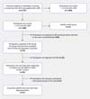
First, the Hypomania Checklist 32 items (HCL-32R)27 was administered to all first-year students at Celal Bayar University (n=2,757). Of these, 1,688 screened positive, defined as a score of = 14 on the HCL-32R. From this group, 1,197 individuals were successfully contacted using the information they had previously provided and agreed to participate in further assessment.
Second, all enrolled participants underwent a phone interview conducted by a psychiatrist using the Mood Disorders Module of Structured Clinical Interview for DSM-IV Axis I disorders (SCID-I).28 This step identified 52 individuals with a possible diagnosis of BD.
Third, these individuals were invited to the Department of Psychiatry at Hafsa Sultan Hospital, Celal Bayar University, where they completed a full SCID-CV interview conducted by a psychiatrist. A diagnosis of BD was confirmed in 30 participants. Of these, 27 met additional inclusion criteria of never having received a formal diagnosis or treatment for BD and agreed to participate in the study. The remaining 22 participants were excluded due to not meeting full diagnostic criteria according to the DSM-IV, or because of diagnoses such as Attention-Deficit/Hyperactivity Disorder (ADHD), personality disorders, or other exclusionary conditions.
Finally, a healthy control group matched for age, sex, and education was selected from participants who screened negative on the HCL-32R. These controls were also assessed using the full SCID-CV interview to confirm the absence of any personal history of psychiatric disorders and any severe psychiatric disorders in their first-degree relatives.
Additional eligibility criteria for all participants were: (i) age between 18 and 30 years; (ii) no history of brain trauma, neurological disorders, or severe medical illness; and (iii) no lifetime history of alcohol or substance abuse or dependence. Patients were excluded if they had a prior diagnosis of any other Axis I or II psychiatric disorder, any history of treatment with antipsychotics or mood stabilizers, any previous psychiatric hospitalization, or a prior diagnosis and treatment for BD.
Clinical assessment
In addition to the diagnostic interviews described above, all participants - regardless of diagnosis - were evaluated for functional outcomes using the Functioning Assessment Short Test (FAST)29 and for psychopathology using the Structured Interview Guide for the Hamilton Depression Rating Scale - Seasonal Affective Disorder Version (SIGH-SAD)30 and the Young Mania Rating Scale (YMRS).31
Cognitive assessment
All participants completed the following cognitive tasks: (i) the Wisconsin Card Sorting Test (WCST), the Stroop Color-Word Test (SCWT), and the Trail Making Test (TMT) to assess executive functions; (ii) the Continuous Performance Test (CPT) to assess sustained attention; (iii) the Digit Span subtest from the Wechsler Adult Intelligence Scale (WAIS) to evaluate working memory; (iv) the Digit Symbol Coding Test (DSCT) from the WAIS as a measure of processing speed, attention, and working memory; and (v) the Rey Auditory Verbal Learning Test (RAVLT) to assess verbal learning and memory.
The Stroop interference effect was calculated using the following formula: color word score - ([word score + color score]/2).32
Assessment of oxidative stress
Peripheral venous blood samples were collected in plain tubes (without anticoagulant) after overnight fasting. Samples were centrifuged at 4,000 rpm for 10 minutes at +4 °C. The separated serum was frozen and stored at -80 °C until analysis. Commercially available assay kits were used to measure SOD, CAT, NO, and TBARS. All analyses were performed in the same laboratory.
One participant from the DNP group was excluded from the oxidative stress analysis due to a spoiled sample.
Neuroimaging
Magnetic resonance imaging (MRI) image acquisition
MRI data for all participants were acquired using the same General Electric Signa HDxt 1.5 Tesla scanner at the Radiology Department of Celal Bayar University. Images were obtained using axial 3D T1-weighted Fast Spoiled Gradient-Recalled Echo (FSPGR) sequences (TE = 7.1-17.0, field of view [FOV] = 24 cm2, slice thickness = 1.6 mm, flip angle = 15°, read bandwidth [RBW] 10.42 kHz, NEX: 1, freq = 256, phase = 192, phase FOV = 0.85).
Image processing
All MRI scans were visually inspected for brain abnormalities and image quality. Cortical thickness and volumetric segmentation were performed using FreeSurfer v5.3.0 (http://surfer.nmr.mgh.harvard.edu/).33-37 Each participant's T1-weighted image was first registered to Talairach space. Images were then segmented into white matter (WM) and gray matter (GM) based on local and neighboring intensity values. The cortical surface of each hemisphere was inflated to an average spherical surface to accurately delineate both the pial surface (GM) and the WM/GM boundary. The cerebral cortex was parcellated into regions according to the Desikan-Killiany atlas.
All regional measures - including cortical thickness and subcortical volumes - were extracted using FreeSurfer. Upon completion, all images were visually inspected again, and any topological inaccuracies were manually corrected.
Statistical analysis
All statistical analyses were performed using IBM SPSS Statistics for Windows, version 21.0. The normality of variable distributions was assessed using the Shapiro-Wilk test. Chi-square tests were used for categorical variables, and Student's t-tests were applied to continuous variables with normal distributions.
Differences in neurocognitive test performance, oxidative stress marker levels, and clinical variables with non-normal distributions were analyzed using Mann-Whitney U tests. The threshold for statistical significance was set at p < 0.008 for demographic and clinical variables, p < 0.002 for cognitive variables, and p < 0.01 for oxidative stress measures, all adjusted using Bonferroni correction for multiple comparisons.
Group differences in cortical thickness were assessed using a general linear model (GLM) implemented in the Query, Design, Estimate, Contrast (QDEC) application of FreeSurfer. Total gray matter volume was included as a covariate in the model. The significance threshold was set at p < 0.01, with Monte Carlo simulation (10,000 iterations) applied to provide cluster-wise correction for multiple comparisons.
Ethics statement
The study protocol was approved by the Regional Ethic Committee of Celal Bayar University, and written informed consent was obtained from all participants prior to their inclusion in the study.
RESULTS
Demographic, clinical, and functional characteristics
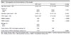
The demographic and clinical characteristics of the two groups are presented in Table 1. One participant from the HC group was excluded due to poor-quality imaging data. There were no significant group differences in age, sex, or education level. Family history of BD and other psychiatric disorders was also similar between groups.
| DNP (n=27) | HC (n=27) | p-value | |
|---|---|---|---|
| Age, years (mean ± SD) | 20.85±2.08 | 21.70±0.99 | 0.0630 |
| Gender (%) | |||
| Female | 59.30 | 59.30 | 1.0000 |
| Male | 40.70 | 40.70 | |
| Education, years (mean ± SD) | 13.33±1.10 | 13.74±1.25 | 0.2100 |
| FAST (mean ± SD) | 18.44±10.97 | 4.74±3.66 | < 0.0001 |
| HAM-D (median) | 3.00 | 1.00 | < 0.0001 |
| YMRS (median) | 3.00 | 0.00 | < 0.0001 |
| BD type (%) | |||
| BD I | 55.60 | ||
| BD II | 44.40 | ||
| Family history of BD in first-degree relatives (%) | 0.00 | 0.00 | |
| Family history of OPD in first-degree relatives (%) | 11.10 | 7.40 | 0.6390 |
BD = bipolar disorder; DNP = drug-naive patients; FAST = Functioning Assessment Short Test; HAM-D = Hamilton Depression Rating Scale; HC = healthy controls; OPD = other psychiatric disorders; YMRS = Young Mania Rating Scale.
None of the DNP participants reported a family history of BD in first-degree relatives; however, one participant reported BD in a second-degree relative. Other psychiatric conditions reported in the DNP group included depressive disorder in the mothers of two participants and ADHD in the brother of one participant. In the HC group, one participant's father and another's mother had been diagnosed with depressive disorder.
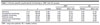
The DNP group showed significantly lower daily psychosocial functioning compared to the HC group across all domains of the FAST (Table 2). At the time of assessment, none of the DNP participants were experiencing a mood episode; however, only 62.9% were asymptomatic (defined as Hamilton Depression Rating Scale [HAM-D] < 8 and YMRS < 5). Although all participants scored below the clinical cutoff of 8 on the HAM-D, 37.03% of the DNP group showed mild manic symptoms, with YMRS scores between 5 and 8, which do not meet the threshold for a manic episode but still reflect subclinical symptoms.
None of the participants in either group reported substance use within the past year.
| DNP | HC | ||||||
|---|---|---|---|---|---|---|---|
| z | U | p-value | Median | Mean rank | Median | Mean rank | |
| Autonomy | -3.19 | 200.50 | 0.001 | 1.00 | 33.57 | 0.00 | 21.43 |
| Occupational functioning | -3.31 | 180.00 | 0.001 | 3.00 | 34.33 | 0.00 | 20.67 |
| Cognitive functioning | -3.89 | 142.50 | 0.000 | 4.00 | 35.72 | 2.00 | 19.28 |
| Financial issues | -4.93 | 90.50 | 0.000 | 3.00 | 37.65 | 0.00 | 17.35 |
| Interpersonal relationships | -5.36 | 63.00 | 0.000 | 3.00 | 38.67 | 0.00 | 16.33 |
| Leisure time | -2.54 | 225.50 | 0.011 | 1.00 | 32.65 | 0.00 | 22.35 |
DNP = drug-naive patients; HC = healthy controls.
Cognitive task performance
The DNP group underperformed on all cognitive tasks compared to the HC group. However, group differences that survived Bonferroni correction were observed in the WCST and RAVLT (Table 3).
| DNP | HC | ||||||
|---|---|---|---|---|---|---|---|
| z | U | p-value | Median | Mean rank | Median | Mean rank | |
| Executive functioning | |||||||
| WCST | |||||||
| Total errors | -4.93 | 80.00 | 0.000 | 26.00 | 38.04 | 17.00 | 16.96 |
| Perseverative responses | -4.31 | 117.00 | 0.000 | 10.16 | 36.67 | 7.81 | 18.33 |
| Categories | -4.43 | 114.00 | 0.000 | 8.00 | 18.22 | 9.00 | 36.78 |
| Stroop task | |||||||
| Word score (s) | -2.39 | 226.00 | 0.017 | 9.64 | 32.63 | 8.58 | 22.37 |
| Color Score (s) | -1.70 | 266.00 | 0.088 | 11.37 | 31.15 | 10.55 | 23.85 |
| Color word score (s) | -2.56 | 216.00 | 0.010 | 22.19 | 33.00 | 17.96 | 22.00 |
| Stroop interference effect┼ | -2.10 | 243.00 | 0.036 | 11.02 | 32.00 | 7.81 | 23.00 |
| TMT-B (s) | -2.65 | 211.00 | 0.008 | 55.84 | 33.19 | 44.52 | 21.81 |
| Working memory | |||||||
| Digit Span Test | |||||||
| Forward | -2.28 | 236.50 | 0.022 | 7.00 | 22.76 | 7.00 | 32.24 |
| Backward | -2.91 | 203.00 | 0.004 | 5.00 | 21.52 | 5.00 | 33.48 |
| Processing speed | |||||||
| TMT-A (s) | -2.58 | 215.00 | 0.010 | 23.46 | 33.04 | 20.84 | 21.96 |
| DSCT | -1.14 | 298.50 | 0.253 | 70.00 | 25.06 | 70.00 | 29.94 |
| Sustained attention | |||||||
| CPT omission error | -2.17 | 262.50 | 0.030 | 0.00 | 31.28 | 0.00 | 23.72 |
| Verbal learning/memory | |||||||
| RAVLT | |||||||
| Immediate recall (total 1-5) | -3.80 | 145.00 | 0.000 | 50.00 | 19.37 | 60.00 | 35.63 |
| Short delay recall (list 6) | -3.41 | 169.00 | 0.001 | 11.00 | 20.26 | 13.00 | 34.74 |
| Long delay recall (list 7) | -3.71 | 151.50 | 0.000 | 10.00 | 19.61 | 13.00 | 35.39 |
| Recognition | -2.16 | 244.50 | 0.030 | 13.00 | 23.06 | 14.00 | 31.94 |
CPT = Continuous Performance Test; DNP = drug-naive patients; DSCT = Digit Symbol Coding Test; HC = healthy controls; RAVLT = Rey Auditory Verbal Learning Test; TMT = Trail Making Test; TMT-A = connecting numbers in ascending order; TMT-B = alternating between numbers and letters in ascending order; WCST = Wisconsin Card Sorting Test.
┼ Interference between language and color information.
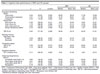
Patients made significantly more total errors and perseverative errors and achieved significantly fewer categories on the WCST. In the RAVLT, patients performed significantly worse across all recall conditions, but the difference in the recognition condition did not reach statistical significance.
Oxidative stress markers
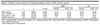
None of the group differences in oxidative stress markers survived correction for multiple testing, except for CAT, which was significantly higher in the DNP group compared to the HC group (Table 4).
| DNP┼ | HC | ||||||
|---|---|---|---|---|---|---|---|
| z | U | p-value | Median | Mean rank | Median | Mean rank | |
| SOD (ng/mL) | -1.26 | 280.00 | 0.206 | 12.73 | 24.27 | 15.80 | 29.63 |
| CAT (nmol/mL) | -2.98 | 183.00 | 0.003 | 66.08 | 33.46 | 46.43 | 20.78 |
| NO (µmol/L) ± SD | -0.64 | 315.00 | 0.522 | 8.85 | 28.38 | 9.52 | 25.67 |
| TBARS (nmol/mL) | -1.53 | 264.50 | 0.124 | 1.31 | 30.33 | 1.14 | 23.80 |
CAT = catalase; DNP = drug-naive patients; HC = healthy controls; NO = nitric oxide; SOD = superoxide dismutase; TBARS = thiobarbituric acid reactive substances.
┼ Data missing one of the patients.
Brain morphometry
Cortical thickness
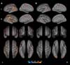
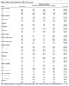
The DNP group exhibited significant cortical thinning in multiple regions compared to the HC group. These included the bilateral temporal lobes, superior and middle frontal gyri, superior and inferior parietal lobules, precentral, postcentral, and paracentral gyri, insula, bilateral precuneus, left orbitofrontal cortex, and the right pars triangularis (Figure 2A; Table 5).

| Talairach coordinates | |||||
|---|---|---|---|---|---|
| Region | Value (log p) | X | Y | Z | Size (mm2) |
| Superior temporal | |||||
| L | 4.24 | -25.7 | 3.8 | 706 | 328.85 |
| R | 3.32 | 47.2 | -29.3 | 7.4 | 371.00 |
| Middle temporal | |||||
| L | 2.98 | -62.3 | -40.3 | -9.6 | 102.13 |
| R | 3.05 | 64.6 | -30.5 | -9.8 | 139.17 |
| Inferior temporal | |||||
| L | 4.21 | -45.1 | -63.9 | -2.1 | 422.63 |
| R | 2.43 | 52.8 | -53.3 | -16.8 | 57.68 |
| Temporal pole | |||||
| L | 2.57 | -35.7 | 14.4 | -37.4 | 80.89 |
| R | 2.17 | 31.0 | 3.2 | -30.1 | 3.78 |
| Precentral | |||||
| L | 3.35 | -48.7 | -8.8 | 40.1 | 94.45 |
| R | 2.85 | 51.0 | 2.9 | 4.7 | 108.70 |
| Postcentral | |||||
| L | 3.56 | -40.1 | -9.4 | 16.3 | 145.87 |
| R | 4.22 | 43.0 | -27.2 | 45.8 | 315.76 |
| Paracentral | |||||
| L | 3.01 | -5.5 | -30.9 | 52.9 | 146.51 |
| R | 2.36 | 7.9 | -20.8 | 57.9 | 45.53 |
| Superior frontal | |||||
| L | 2.67 | -20.7 | 24.2 | 46.4 | 122.93 |
| R | 2.66 | 8.7 | 0.7 | 56.4 | 123.43 |
| Rostral middle frontal | |||||
| L | 2.30 | -17.4 | 49.7 | 26.7 | 26.13 |
| R | 3.67 | 38.3 | 46.1 | 20.6 | 300.94 |
| Caudal middle frontal | |||||
| L | 3.31 | -27.1 | 0.7 | 47.6 | 129.49 |
| R | 3.52 | 29.4 | 17.0 | 44.2 | 106.06 |
| Superior parietal | |||||
| L | 2.36 | -28.1 | -63.8 | 40.0 | 52.56 |
| R | 2.94 | 24.6 | -56.7 | 59.2 | 224.23 |
| Inferior parietal | |||||
| L | 4.18 | -39.9 | -53.0 | 17.1 | 311.34 |
| R | 2.81 | 44.5 | -62.5 | 35.4 | 194.16 |
| Supramarginal | |||||
| L | 2.74 | -60.6 | -28.0 | 27.2 | 183.41 |
| R | 4.93 | 60.0 | -28.1 | 36.9 | 819.24 |
| Precuneus | |||||
| L | 2.54 | -5.8 | -69.7 | 41.2 | 53.32 |
| R | 2.17 | 5.7 | -60.0 | 18.7 | 22.46 |
| Posterior cingulate | |||||
| L | 2.72 | -5.7 | -7.7 | 40.2 | 80.58 |
| R | 2.63 | 4.5 | -5.6 | 36.7 | 20.88 |
| Isthmus cingulate | |||||
| L | 2.19 | -4.3 | -37.3 | 31.4 | 15.25 |
| R | 2.21 | 4.7 | -39.1 | 31.2 | 19.11 |
| Insula | |||||
| L | 2.18 | -36.6 | 0.6 | -8.9 | 15.67 |
| R | 3.51 | 34.6 | -23.1 | 20.7 | 193.23 |
| Medial orbitofrontal | |||||
| L | 3.56 | -5.1 | 53.5 | -20.5 | 91.59 |
| Lateral orbitofrontal | |||||
| L | 2.44 | 16.0 | 43.1 | -18.1 | 43.47 |
| Pars opercularis | |||||
| L | 2.63 | -50.6 | 11.4 | 3.9 | 114.24 |
| Pars triangularis | |||||
| R | 2.47 | 44.4 | 37.7 | 0.3 | 58.65 |
| Fusiform | |||||
| L | 2.48 | -16.0 | 43.1 | -18.1 | 43.47 |
HC = healthy controls (n=26; one excluded due to poor image quality); DNP = drug-naive patients with bipolar disorder (n=27). p < 0.01.
L = left hemisphere; R = right hemisphere.
DISCUSSION
In the present study, psychosocial functioning, neurocognitive performance, oxidative stress, and brain structure were examined in a sample of students with previously undiagnosed and untreated BD.
Among the 1,197 respondents who screened positive on the HCL-32, we identified 30 cases of previously undiagnosed BD, yielding a point prevalence of 2.5%, assuming a similar rate among those who screened positive (n = 491) but did not participate in the subsequent assessments. This prevalence rate is remarkably close to the 2.4% reported in the World Mental Health Survey (WMH).2
Although the patients identified in this study could be considered high-functioning, as they were university students, their psychosocial functioning - specifically in the domains of autonomy, occupational functioning, cognitive functioning, financial issues, interpersonal relationships, and leisure time, as assessed by the FAST - was significantly lower than that of their peers without mental health problems. These findings are consistent with those reported in more chronic, euthymic patients with BD.38,39 Furthermore, they confirm that these impairments are independent of psychotropic treatment, as the current sample had never received medication.40,41 Notably, to our knowledge, this is the first study to examine psychosocial functioning in never-medicated patients with BD.
Previous studies have reported that impaired psychosocial functioning is associated with neurocognitive dysfunction and subthreshold affective symptoms.14,15,42 Although only seven participants in the DNP group exhibited subthreshold manic symptoms, these symptoms were very mild, with a maximum YMRS score of 8, yet still higher than those reported by the HC group. This finding may suggest that a persistent vulnerability to affective morbidity is present in patients even outside of full-blown mood episodes. Such vulnerability may negatively impact their engagement in social roles and activities.
An extensive body of research has shown that many patients with BD exhibit significant cognitive impairments, regardless of mood state, particularly in executive function, attention, verbal learning, and memory.8,11,43 In the present study, patients with BD underperformed across all cognitive tasks; however, statistically significant differences were observed in the WCST and RAVLT, which primarily assess abstraction/cognitive flexibility and verbal learning and memory, respectively. Abstraction and cognitive flexibility are typically mediated by the dorsal and ventral prefrontal cortices. Nevertheless, previous studies have demonstrated that successful performance on the WCST relies on the engagement of a widespread neural network, involving prefrontal, frontal, temporal, parietotemporal, and parieto-occipital cortical regions during various stages of the task.44,45
Furthermore, one study demonstrated a correlation between the recognition of familiar words and activation in the right lateral prefrontal cortex as well as between the processing of novel words and the right anterior hippocampus.46
The cognitive profile observed in the current sample aligns with the neuroimaging findings, which revealed cortical thinning in several regions, including the prefrontal and temporal cortices, areas critically involved in cognitive control and verbal learning and memory. Structural abnormalities in these regions, among others, have also been reported in the largest case-control study of BD to date, conducted by the Enhancing Neuro Imaging Genetics through Meta-Analysis (ENIGMA) Consortium, which included data from 2,447 patients.17 The present findings further support the notion that such structural brain abnormalities are an integral component of syndromal expression of BD.
Oxidative stress, along with associated lipid peroxidation, has been proposed as a potential mechanism underlying brain abnormalities in BD, thereby contributing to the manifestation of clinical symptoms and cognitive deficits. Two meta-analyses have reported significantly elevated levels of NO and TBARS in blood cells or serum from patients with BD, without concomitant changes in antioxidant enzymes (e.g., SOD, CAT, and GPx).18,21
However, our findings suggest that oxidative stress may not be present in the early stages of the disease but may instead emerge in more severe cases or as a consequence of illness progression. In contrast to the expected pattern of oxidative imbalance, patients in this study showed a significant increase in CAT levels. This is consistent with a prior study in early-stage BD, which also reported elevated CAT levels.47 Additionally, another study found a negative correlation between CAT levels and illness duration in more chronic patients.48
Another possible explanation is that the oxidative stress markers used in this study are not sensitive enough to detect early alterations. For example, increases in protein carbonyl content (PCC), a marker of oxidative protein modification, have been detected in population-based samples that identified patients with less severe or early-stage presentations, similar to the current study.49
Moreover, elevated TBARS levels appear to be primarily observed in studies involving chronic patients or those with more severe clinical presentations. Accordingly, previous research found no case-control differences in TBARS in early-stage BD47 or in population-based studies of BD with a milder disease course.49 Similarly, Siwek et al.50 reported that TBARS levels were increased only in symptomatic patients, thereby suggesting lipid peroxidation becomes more prominent as the number of mood episodes accumulates over time.
The main strength of the present study lies in the inclusion of a sample of high-functioning, early-stage, never-medicated patients with BD who were not experiencing a mood episode at the time of assessment. Consequently, the findings are free from confounding factors associated with chronicity, psychotropic medication, and acute psychopathology. Patients were carefully matched to controls in terms of academic achievement and education, as the entire sample consisted of university students. This matching strategy helps mitigate potential case-control differences that could otherwise be attributed to the effects of educational attainment. Additionally, the study adopted an epidemiological approach, rather than focusing on help-seeking or clinically referred populations. Such designs enhance the representativeness of the sample, albeit within a population of high-achieving individuals. Finally, both patients and controls underwent a comprehensive assessment, including molecular, brain structural, cognitive, and functional evaluations. This multimodal approach provides a more comprehensive understanding of the neurobiology of BD than is typically achieved in most studies. Although the sample size is relatively small, this is consistent with the generally low prevalence of BD in population-based samples and aligns with point prevalence estimates reported in the literature. The cross-sectional nature of the study does not permit conclusions about the progression of the detected abnormalities, but it provides a critical foundation for future longitudinal studies aimed at tracking the clinical trajectories of these patients and their associations with cognition, brain structure, and oxidative stress.
In summary, this study is the first to demonstrate that cognitive, functional, and brain structural alterations are already present in the early stages of BD, even among high-achieving individuals. These findings contribute valuable insights to the current understanding of the neurobiology of BD and advance the existing body of knowledge on the disorder.
ACKNOWLEDGEMENTS
This study was supported by the Incentive Award for Research Project of the Turkish Psychiatric Association; 18th Annual Meeting and Clinical Education Symposium of the Turkish Psychiatric Association, April 2014, Antalya, Turkey. Research Award of the Turkish Psychiatric Association; 52nd National Psychiatry Congress of Turkish Psychiatric Association, November 2016, Antalya, Turkey.
 PDF
 Print
 Send this article by email
 How to cite this article
 Submit a comment
 Mendeley
 Pocket
 Share on Facebook
 Share on Twitter