



ABSTRACT:
OBJECTIVE: Depressive symptoms during the perinatal period significantly impact mothers and infants.
Emerging evidence suggests a connection between gut microbiota and mood regulation.
This study investigated whether depressive symptoms are associated with changes in
the gut microbiota of women during the perinatal period.
METHODS: Thirty-four pregnant women were screened for depression using the Edinburgh Postnatal
Depression Scale (EPDS) and categorized based on symptom severity. Stool samples were
collected during the third trimester and at two postpartum timepoints. All samples
underwent 16S rRNA gene sequencing and quantification of short-chain fatty acids (SCFA)
by gas chromatography-mass spectrometry (GC-MS).
RESULTS: No differences in SCFA concentrations were observed between groups (p > 0.05). However,
postpartum women with moderate to severe symptoms (MS group) had a significant increase
in Enterobacteriaceae abundance compared to women with mild or absent symptoms (AM
group) (p < 0.05). The Bifidobacterium genus increased significantly in both groups over time (p < 0.05). The MS group showed
a reduction in depressive symptoms during psychiatric treatment (p < 0.05).
CONCLUSION: These findings suggest a link between gut microbiota and perinatal depressive symptoms.
Further research using microbiome-targeted approaches is needed to understand the
broader implications for maternal health.
Keywords: Gut-brain axis; perinatal; microbiome; depression.
FIGURES
| Note: How to cite this article: Mota AS, Sparvoli LG, Vanzele PAR, Naspolini NF, Tobaruela EC, Yoshizaki CT, et al. Longitudinal gut microbiota composition during the perinatal period in women with different intensities of depressive symptoms. Braz J Psychiatry. 2025;47:e20243721. http://doi.org/10.47626/1516-4446-2024-3721 |
| Received: May 09 2024; Accepted: August 16 2024 |
INTRODUCTION
Pregnancy and the postpartum period are described as a time of physical and psychological transformation. These changes can lead to significant mood alterations, which may worsen without network support from the community and healthcare professionals. A lack of early diagnosis and absence of familial and financial support further exacerbates this psychological state, making women more vulnerable and facilitating the onset of a mental disorder such as perinatal and postpartum depression.1,2
Poor maternal health can directly impact fetal neurodevelopment. For example, depression during this period can result in consequences such as premature birth and low birth weight, compromised psychological and intellectual child development, onset of behavioral and psychiatric disorders, muscle loss, and increased risk of diarrhea and respiratory illnesses.1,3-5
One of the recommended methods for screening depression during gestation is the application of standardized tests, such as the self-administered Edinburgh Postnatal Depression Scale (EPDS). Despite not being a diagnostic method, the EPDS has demonstrated high specificity and sensitivity for screening depressive symptoms, however, this method still has a very low implementation in prenatal appointments.1,6,7
It is well established that women with depressive symptoms may experience an increase in cortisol levels, which can lead to immune system disruption, consequently affecting fetal blood circulation and promoting a higher concentration of inflammatory cytokines capable of causing disturbances in fetal neurodevelopment. Furthermore, these heightened cortisol levels can also disturb gut barrier permeability and stimulate the activation of toll-like receptors, triggered by pathogen-associated molecular patterns (PAMPs).8-12
Recently, the gut microbiota has been associated with psychiatric disorders, including depression, through alterations in the gut-brain axis. An increase in pro-inflammatory members of the Enterobacteriaceae family and a decrease in quantification of short-chain fatty acids (SCFA)-producing bacteria, such as the genus Bifidobacterium, have been found in the gut microbiota of women diagnosed with depression.13,14 In addition, the composition of the intestinal microbiota undergoes significant changes during pregnancy, varying across the three trimesters but most markedly in the first and third. In the third trimester, healthy pregnant women demonstrate an increased abundance of Bacteroides and Akkermansia genera, both indicative of a eubiotic microbiota. The onset of diseases, especially in the final trimester, appears to be linked to changes in microbiota composition. This is evidenced by a decrease in anti-inflammatory-related microbes such as Faecalibacterium and an increase in abundance of the Lachnospiraceae, genus Phascolarctobacterium, and Christensenellaceae. Furthermore, the microbiotic profile of women with high weight gain in the third trimester of pregnancy resembles that of obese individuals, characterized by an elevated abundance of the Actinomycetota and Pseudomonadota phyla.15-17
Kelsey et al.3 found an association between maternal microbiota composition and the behavior of newborns. They also demonstrated that associated virulence factors enabled colonization by pathobionts, which was associated with increased connectivity in the neural network associated with negative emotions. These findings are supported by Zhang et al.,18 who observed that offspring of rats treated with antibiotics during pregnancy exhibited reduced sociability. Although the exact mechanism responsible for behavioral changes related to maternal-fetal interaction and the vertical transmission of microbiota remains unclear, it is acknowledged that a connection between maternal intestinal composition and neurodevelopment exists.
Within this context, we aim to investigate whether different intensities of depressive symptoms are associated with changes in the gut microbiota of women during pregnancy and the postpartum period.
METHODS
Study design and participants
A cohort of 34 pregnant women were recruited from Hospital das Clínicas da Faculdade de Medicina da Universidade de São Paulo (HCFMUSP) and Hospital Universitário (HU-USP). The inclusion criteria were 1) third trimester of pregnancy; 2) age 18-45 years; 3) receiving prenatal care at HCFMUSP or HU-USP. The exclusion criteria were: 1) cognitive dysfunction or chronic infection; 2) diagnosis of fetal malformation incompatible with life; 3) psychiatric disorders other than depression; 4) use of antibiotics. All participants were informed about the study and provided consent. This study was approved by the ethics committees of HCFMUSP, HU-USP, and Faculdade de Ciências Farmacêuticas (FCF-USP; reference number CAAE 22725119130030076).
Data collection
Questionnaires were used to collect sociodemographic data, dietary information, and clinical history, including a history of depression (defined as a diagnosis of depression made by a psychiatrist prior to the current pregnancy). The EPDS was used as a screening tool to allocate the participants into groups in accordance with presence and severity of depressive symptoms: absent or mild (AM) (n=16) and moderate or severe (MS) (n=18). Because participants from the AM group had no indications for treatment, they did not receive antidepressants. Only participants from the MS group received psychiatric care, which included administration of antidepressants, during pregnancy and postpartum. Exposure to antidepressants was included as a covariate.
Maternal stool samples were collected at three different time points: the first (G) was during the third trimester of gestation (28-40 weeks); the second (P1), during postpartum hospitalization (between postpartum days 1 and 3); and the third (P2), at the 1-month postpartum follow-up appointment. The samples were frozen at -20 °C for 24 hours maximum for subsequent storage at -80 °C until processing.
Assessment of depressive symptoms
The EPDS is a self-administered scale comprising 10 questions, each with four possible answer choices; each answer can be scored from 0 to 3 points. The maximum score is 30, with higher scores denoting more severe depressive symptoms. Participants completed the EPDS at enrollment and at each sample collection. A cutoff of = 10 was used to classify moderate to severe symptoms, while scores below that threshold were categorized as absent or mild depressive symptoms.19
DNA extraction and sample sequencing.
DNA from the stool samples was extracted using the QIAamp PowerFecal Pro DNA Kit (QIAGEN, Hilden, Germany), following the manufacturer's recommendations. The DNA was quantified in Qubit equipment (Thermo Fisher Scientific, Waltham, USA).
Following DNA extraction, amplification of the V4 region of the bacterial 16S ribosomal segment was performed as previously described by Kozich et al.20 The protocol followed for this region involved the use of primer sequences according to the standard International Union of Pure and Applied Chemistry (IUPAC) nucleotide nomenclature, which were as follows:
16S forward primer sequence, 5´-TCGTCGGCAGCGTCAGATGTGTATAAGAGACAGCCTACGGGNGGCWGCAG-3´; 16S reverse primer sequence, 5´-GTCTCGTGGGC TCGGAGATGTGTATAAGAGACAGGACTACHVGGGTATCTAATCC-3´.
All steps were carried out following the manufacturer's instructions (Illumina-16S Metagenomic Sequencing Library Preparation).21 After obtaining the sequences, sample demultiplexing was performed based on the corresponding barcode sequences.
Short-chain fatty acids detection and quantification
The quantification of SCFAs (acetate, propionate, and butyrate) was performed at FCF-USP, in collaboration with the Bioactive Compounds Research Group. SCFA extraction was carried out using water acidified with 0.5% phosphoric acid and ethyl acetate with the addition of an internal standard (2-methylpentanoic acid). The analysis was conducted on a gas chromatograph-mass spectrometer (GC-MS) (Agilent 5977, Carpinteria, CA, USA) under optimized conditions.
Chromatographic conditions
A 1-µL sample was injected in splitless mode with an injector temperature of 240 °C. Helium was used as the carrier gas at a constant flow rate of 2.9 mL/min. The chromatographic column used was CPWax (30 m × 0.32 µm × 0.2 µm). The temperature program started with a ramp at 15 °C/min from 50 °C to 180 °C, followed by an increase of 35 °C/min to 200 °C, and then 50 °C/min to 250 °C. The chromatograph/mass spectrometer interface was operated at a temperature of 250 °C. Electron impact ionization (70 eV) was used with the ion source held at 230 °C. The mass range was set from 70-600 m/z at a scan rate of 20 scans/s.
Bioinformatic and statistical analysis
Quantitative Insights into Microbial Ecology (QIIME 2.0) was used to analyze the 16S rRNA library results obtained through sequencing. The sequences from the resulting libraries were clustered into ASVs to identify 97% similarity based on the Silva database, version 128.
Statistical analysis was conducted using R software (version 4.2.2). The obtained data were tested for normality using the Shapiro-Wilk method. For variables with non-normal and non-homogeneous distribution, the non-parametric Mann-Whitney or Wilcoxon test was employed. Pearson's chi-square test or Fisher's exact test was used to assess associations between two categorical variables. Regression analysis was performed using the MAaslin2 package, and correlation analysis was conducted using the Spearman test. For paired samples with a non-normal distribution, the Friedman test with post-hoc Dunn procedure was used. For beta diversity analysis, the permutational multivariate analysis of variance (PERMANOVA) test was applied, and alpha diversity metrics (Shannon, Chao1, and Simpson) were analyzed using the Wilcoxon test, with the p-value adjusted using the Benjamini-Hochberg (BH) procedure. The significance level was set at p = 0.05.
We used a linear mixed-effects model (nlme R package22) with repeated measures to evaluate the longitudinal relationship between groups and the Bifidobacterium genus and Enterobacteriaceae family. This analysis considered both the presence and absence of the interaction term between timepoints and groups.
RESULTS
Sociodemographic characteristics
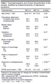
The average age range was 30-35 years and did not differ between groups (p = 0.35). Educational attainment (p = 0.13) and marital status (p = 0.59) were also similar. The MS group presented higher EPDS scores (14.1 ± 4.2) compared to the AM group (4.8 ± 2.6; p < 0.001) (Table 1). Participants within the MS group had a higher parity (p = 0.01), history of miscarriage (p = 0.01), and psychiatric history before pregnancy (p < 0.001). Only women diagnosed with depression received medication. Regarding missing data, one participant in the AM group did not select any of the education alternatives, and one from the MS group did not respond regarding the history of miscarriage. Because symptoms of depression were, by definition, more severe in the MS group, all participants from this group were followed by a mental health team and received antidepressant pharmacotherapy throughout pregnancy and the postpartum period.
| Characteristics | AM (%) | MS (%) | p-value |
|---|---|---|---|
| Age (mean ± SD) | 30.9±5.4 | 32.7±6.2 | 0.35 |
| Educational attainment | 0.135 | ||
| Complete high school | 46.7 | 60.6 | |
| Undergraduate | 53.3 | 27.8 | |
| Marital status | 0.59 | ||
| Single | 25.0 | 33.3 | |
| Married | 75.0 | 66.7 | |
| Gestation | 0.01* | ||
| Primiparous | 68.8 | 27.8 | |
| Multiparous | 31.2 | 72.2 | |
| Mode of delivery | 0.02** | ||
| Cesarean | 25.0 | 88.9 | |
| Vaginal | 75.0 | 11.1 | |
| History of miscarriage | 0.01* | ||
| No | 93.8 | 58.8 | |
| Yes | 6.2 | 41.2 | |
| History of depression┼ | < 0.001*** | ||
| No | 93.8 | 27.8 | |
| Yes | 6.2 | 72.2 | |
| Exposure to antidepressants | < 0.001*** | ||
| No | 100.0 | 0.0 | |
| Yes | 0.0 | 100.0 | |
| EPDS score (mean ± SD) | 4.8±2.6 | 14.1±4.2 | < 0.001*** |
Microbiota composition in the third trimester of pregnancy
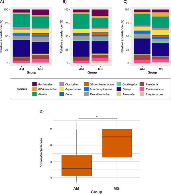
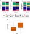
Although both groups had the same predominant bacterial genera, their relative abundances differed - especially regarding the Enterobacteriaceae family, with a relative abundance of 3.2% in the MS group and 0.4% in the AM group. However, this difference was not statistically significant (p = 0.11). Both groups had Blautia as the most abundant genus (21.8% in the MS group and 24.2% in the AM group); however, some genera stood out due to the difference in their average abundance between the groups, as evidenced by the Bacteroides relative abundance of 19.5% in the MS group versus 5.3% in the AM group and the Enterobacteriaceae family, which appeared to be more abundant in the MS group (3.2%) than in the AM group (0.4%), although again the difference was not significant. Other genera were notably increased, such as the genus Oscillospira, which was twice as abundant in the MS group than in the AM group (1.6% versus 0.8%) and the genus Bifidobacterium, which, conversely, was more abundant in the AM group (3.2% versus 2.2% in the MS group). Bacterial genera with < 1% relative abundance were labeled as "Others" and were slightly increased in the AM group, which may indicate an increase in microbial diversity. Nevertheless, none of these findings reached statistical significance (all p > 0.05), either for abundance or alfa and beta diversity (Supplementary Figure S1). The composition of the gut microbiota in women in the third trimester of pregnancy is illustrated in Figure 1A.
Microbiota composition in the post-partum
The abundance of the Enterobacteriaceae family at P1 was increased in the MS group (4.9%) compared to the AM group (1%; p < 0.05) as illustrated in Figures 1B and 1D. This difference in abundance was not reflected in the community structure analyzed in alpha and beta diversity indices (Supplementary Figure S2). Blautia was the most abundant in both study groups. Similar to the relative abundance observed in the 3rd trimester's samples, Bacteroides exhibited greater relative abundance in the MS group (11.8%) compared to the AM group (5.7%), whereas Faecalibacterium showed higher abundance in the AM group (8.2%) compared to the MS group (6.7%). Although these differences were not statistically significant (p > 0.05).
At 1 month postpartum (P2), we observed a trend towards an increase in the relative abundance of the genus Bifidobacterium in the MS group. Additionally, the relative abundance of the Enterobacteriaceae family was more pronounced in the MS group (2.4%), while in the AM group, this family had 0.03% of relative abundance (Figure 1C). However, these findings did not achieve statistical significance except for the abundance of Enterobacteriaceae at P1 (Figure 1D).
Short-chain fatty acids concentration between groups
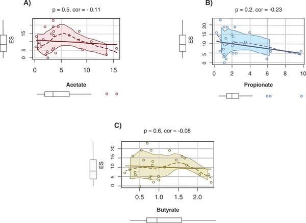
We found no difference in the concentration of SCFAs between the groups, whether in the perinatal or postpartum periods. In addition, no correlation was found between SCFAs and EPDS scores (acetate, p = 0.5, cor = -0.11; propionate, p = 0.2, cor = -0.23; butyrate, p = 0.6, cor = -0.08), as illustrated in Figures 2A, 2B, and 2C, respectively.
Reduction of depressive symptoms and changes in gut microbiota composition during psychiatric treatment
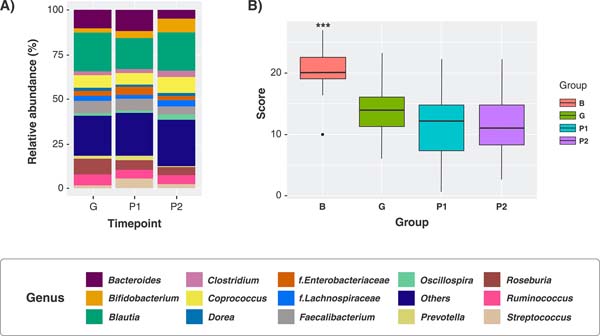
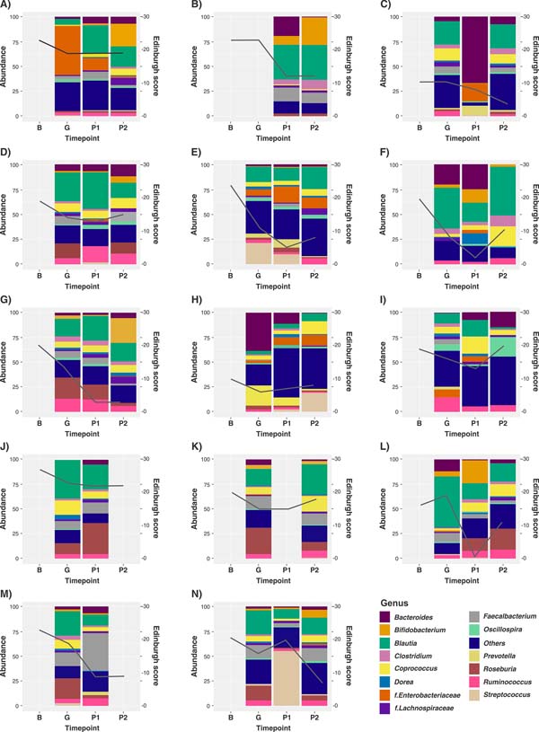
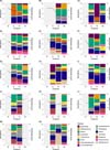
The microbiota composition in the MS group across time is illustrated in Figure 3A. Regarding EPDS scores in the MS group, we observed a significant decrease in depressive symptoms over time (p < 0.05), as treatment progressed (Figure 3B). The Bifidobacterium genus did not show any significant differences when the analysis was carried out cross-sectionally.
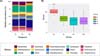
Longitudinal evaluation of gut microbiota across time during perinatal period
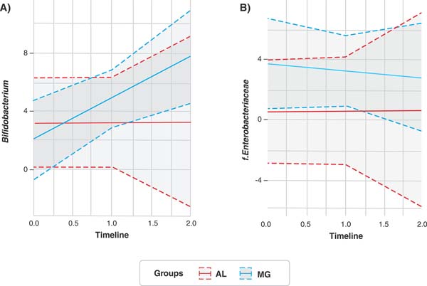
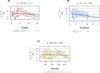
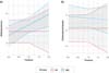
For longitudinal analysis of the Bifidobacterium genus, the combined effect of groups and time yielded an increase of Bifidobacterium within the MS group (ß = 2.84; p = 0.01) but not within the AM group (ß = 0.04; p = 0.97) (Figure 4A). For longitudinal analysis of the Enterobacteriaceae family, the combined effect of groups and time resulted in no difference within either the MS group (ß = -0.46; p = 0.69) or the AM group (ß = 0.06; p = 0.97), as illustrated in Figure 4B.
We also observed an inverse, though not statistically significant, correlation between Bifidobacterium and the Enterobacteriaceae family (cor = -0.14, p = 0.2) (Supplementary Figure S3).
Composition of gut microbiota of participants of MS group across time
In the MS group, the EPDS score decreased over time, reflecting a longitudinal observation of the treatment of depression. While no specific bacterial genus was directly linked, a notable correlation was observed between fluctuations in EPDS scores and the augmentation of certain beneficial bacterial genera. For instance, a correlation between increasing Bifidobacterium abundance and decreasing EPDS score (p = 0.2, cor = -0.19), as shown in Figures 5A, 5B, 5G, 5P, and Supplementary Figure S3, corresponded to a concurrent maintenance of EPDS scores, alongside a noticeable rise in Enterobacteriaceae family abundance (Figures 5A and 5H).
DISCUSSION
Our study shows that the gut microbiome of pregnant women with different intensities of depressive symptoms during the third trimester and postpartum can vary, especially regarding relative abundance of the Enterobacteriaceae family.
Overall, women in the MS group showed an increase in Bifidobacterium abundance and a decrease in Enterobacteriaceae, along with a significant improvement in depressive symptoms during treatment. Moreover, there was a trend toward increasing abundance of the Bifidobacterium genus across time in the MS group under psychiatric treatment. Huang et al.23 also observed an increase in the Actinobacteria phylum after ketamine treatment in animals subjected to experimental depression, suggesting that this phylum may be a potential biomarker for the effectiveness of antidepressant treatment. Although the mechanisms behind these hypotheses are not fully understood, it is known that one of the most abundant genera within the Actinobacteria phylum, Bifidobacterium, produces acetate. This is in line with our findings, as we also identified that acetate concentrations correlated inversely with the severity of depressive symptoms, although not significantly. Some species of the Bifidobacterium genus are associated with serotonin (5-HT) biosynthesis through the modulation of enterochromaffin cells, increased levels of brain-derived neurotrophic factor (BDNF), and reduced cortisol levels; they also influence microglial maturation through gene regulation.24,25 However, we did not find any significant correlation between acetate concentrations and the Bifidobacterium genus in our study, nor did we find statistically significant differences in the concentration of other SCFAs between the AM and MS groups or over the course of treatment within the MS group. This suggests that the trend toward an increase in Bifidobacterium is not reflected in SCFA production, and the increase in SCFAs correlated with the decrease in depressive symptoms may involve other genera and pathways of the gut-brain axis. The limited number of samples could also have made the present study underpowered to detect significance in this association.
The Enterobacteriaceae family is comprised of gram-negative bacteria with disbiotic potential; these bacteria can translocate to the circulatory and lymphatic systems, promoting an increase in the immune response and the release of inflammatory cytokines that can cross the blood-brain barrier and lead to neuroinflammation, a common condition in women with depression.26,27 Fang et al.8 was able to correlate cytokine concentration in plasma with depressive symptomatology during pregnancy. We observed a significant increase in the abundance of Enterobacteriaceae in the MS group, especially at postpartum time point P1, and a notable decrease in its abundance at P2. Interestingly, this decrease mirrors the progressive increase of the Bifidobacterium genus, although not significantly. This is likely due to the production of acetate by Bifidobacterium, an SCFA that is essential for protection against pathogenic bacteria such as Escherichia-Shigella, a genus belonging to the Enterobacteriaceae family (which is significantly increased in the MS group).
The presence of bacteria capable of generating inflammatory responses such as members of the Enterobacteriaceae family appears to be increased in the MS group compared to the AM group. This profile changes with the progression of psychiatric treatment and the improvement of depressive symptoms. The increase in the Clostridium genus and the decrease in the Oscillospira and Prevotella genera may be associated with an increase in the production of SCFAs, which are anti-inflammatory microbes due to their action in suppressing histone deacetylase.28
Regarding taxonomic diversity, previous studies do not provide a consensus on depression-related changes in microbiome alpha diversity. We did not find significant differences in alpha diversity between the AM and MS groups in this cohort. Liu et al.13 reported that more than half of the studies involving microbiota and depression did not show significant differences between groups in terms of alpha diversity, which is consistent with our results.
Regarding EPDS scores, the observed variations indicate that women's treatment experiences are individualized, with some demonstrating rapid, effective responses to treatment while others are slower to respond, requiring additional medication. This variability is noteworthy when examining fluctuations in microbiota, as there is evidence suggesting that, when EPDS scores remain stable in certain women, their corresponding microbiota compositions also remain relatively unchanged, albeit with minor fluctuations in the relative abundance of genera. Conversely, notable shifts in EPDS scores often coincided with more pronounced variations in Bifidobacterium abundance.
Potential limitations of our study should be considered. The sample size was not sufficient to ensure high statistical power, and some potential differences did not reach statistical significance. Strengths of our study include the use of longitudinal microbiome data combined with the assessment of SCFA in pregnant women stratified by presence and severity of maternal depressive symptoms. To our knowledge, this is one of the first studies to characterize the gut microbiota in this population. Finally, this study has the potential to inform preventative intervention strategies to address depression during pregnancy in prenatal care.
Our findings highlight a notable decline in depressive symptoms from baseline to the P2 period, underscoring the importance of integrating EPDS screening into prenatal care and of considering the Enterobacteriaceae family as a plausible biomarker of (or contributing factor in) the onset of maternal depressive symptoms during late pregnancy and postpartum recovery. EPDS scores seem to undergo variations that can be reflected in the microbiota of women with moderate-to-severe depressive symptoms. Further investigation into the precise mechanisms governing these shifts in microbiome composition and their potential impact on maternal mental health is needed. Additionally, comprehending the enduring effects of these microbial variations on maternal and infant health outcomes is crucial for devising targeted interventions and support strategies aimed at enhancing the well-being of pregnant women and their offspring. These findings hint at a potential correlation between microbiota composition and depressive symptoms.
ACKNOWLEDGEMENTS
This study was supported by Coordenação de Aperfeiçoamento de Pessoal de Nível Superior (process 88887.363685/2019-00), Conselho Nacional de Desenvolvimento Científico e Tecnológico, Fundação de Amparo à Pesquisa do Estado de São Paulo (2023/0617/4).
 PDF
 Print
 Send this article by email
 How to cite this article
 Submit a comment
 Mendeley
 Pocket
 Share on Facebook
 Share on Twitter